Objective. In this prospective
randomized study, fetal behavior was
investigated in order to determine the standard
parameters of fetal movements and facial
expressions in all three trimesters of normal
pregnancy.Methods. Sixty-three pregnant women
with singleton pregnancies in all trimesters
were included in the investigation.
Four-dimensional (4D) ultrasound was performed
for each patient over a 30-minute period.
Variables of maternal and fetal characteristics
including gestational age, eight fetal movement
patterns in the first trimester, and sixteen
parameters of fetal movement and fetal facial
expression patterns in the second and third
trimesters were recorded for the construction of
fetal neurological charts.
Results. In the first trimester, a
tendency towards an increased frequency of fetal
movement patterns with increasing gestational
age was noticed. Only the startle movement
pattern seemed to occur stagnantly during the
first trimester (p > 0.05). At the beginning
of the second trimester, the frequency of fetal
movement patterns tended to increase. During the
second and third trimester, multiple regression
and polynomial regression revealed statistically
significant changes in tongue expulsion (p <
0.05), smiling (p < 0.05), grimacing (p <
0.05), swallowing (p < 0.05), eye blinking (p
< 0.01), head movements, and all hand to body
contact movements (p < 0.01), except for head
anteflexion (p > 0.05).
There were no statistically significant
changes during the second and third trimesters
in mouthing, yawning, and sucking (p >
0.05). At the middle of the third trimester, the
fetuses displayed decreasing or stagnant
incidence of fetal facial expressions except for
eye blinking, which showed increased frequency
with increasing gestational age. A statistically
significant correlation was found between all
head movements and hand to body contact patterns
during the second and third trimesters except
for head anteflexion (r = -0.231; p >
0.05).
Conclusions. The full range of
quantitative fetal facial expressions and fetal
movement patterns can be assessed successfully
by 4D sonography. It is important to be able to
assess normal fetal behavior throughout
gestation to identify abnormal behavior before
birth
Introduction
Understanding the structure and function of
the fetal nervous system has been a dream of
physicians for centuries. The number of studies
showing that many neurological problems, such as
minimal cerebral dysfunction, schizophrenia,
epilepsy, and autism, result at least in part
from prenatal neurodevelopmental problems is
increasing.
Fetal behavior can be defined as any
observable action or reaction (to an external
stimulus) by the fetus. This may be recorded by
maternal perception of movement or real-time
ultrasound imaging by means of which fetal
behavior can be observed in the clearest and
most detailed way. Innovations in ultrasonic
technology have created new possibilities in the
study of feta behavior. The introduction of
four-dimensional ultrasound (4D US) has led to
very important conclusions concerning fetal
behavior by enabling us to produce measurable
parameters for the assessment of normal
neurobehavioral development . It is now possible
to study a full range of facial expressions
including smiling, crying, scowling, and eyelid
movements in almost real-time by 4D US.
Analysis of the dynamics of fetal behavior
has led to the conclusion that fetal behavioral
patterns directly reflect developmental and
maturational processes of the fetal central
nervous system. As we learned from postnatal
studies of neonatal behavior, assessment of
behavior is a better predictor of
neurodevelopmental disability than neurological
examination. These findings implicated that
understanding the relation between fetal
behavior and developmental processes in
different periods of gestation would make
possible the distinction between normal and
abnormal brain development, as well as the early
diagnosis of various structural or functional
abnormalities.
The preliminary results of multicentric
studies of fetal brain function suggest that the
study of fetal behavior should be standardized
as much as possible. If behavioral analysis is
to have a role in the routine clinical
environment, then normal standard parameters and
objective methods need to be developed. De Vries
and colleagues were the first to provide a
systematic and detailed classification and
quantitative longitudinal analysi of fetal
behavior during the first half of pregnancy
using two-dimensional ultrasound (2D US).
During the past three years, Kurjak et al.
have initiated extensive research into fetal
behavior in normal and pathological pregnancies
by both threedimensional (3D US) and
four-dimensional ultrasound (4D US).
This study reports reference ranges with
gestational age for suggested use as fetal
neurobehavioral development parameters in normal
singleton pregnancies. Standard movement pattern
and facial expression pattern curves have been
constructed for all trimesters of
pregnancy.
In the first trimester using 4D US one can
simultaneously assess movements of the fetal
head, body, and all fou extremities in three
dimensions. Therefore, the earliest phases of
the human anatomical and motor development can
be visualized and studied simultaneously . It is
possible to study total fetal facial activities
by 4D US. In addition to yawning,
sucking, and swallowing described by 2D
real-time imaging, it is now possible to study a
full range of facial expressions including
smiling, crying, and eyelid movements with this
technology.
Furthermore, four-dimensional sonography
seems to be the method of choice for detecting
subtle changes such as superimposed rotations
and changes in direction of the movements. The
first spontaneous fetal movements can be
observed at postconceptional weeks 7 to 7.5. In
the subsequent weeks (8th to 9th weeks of
gestation), they are replaced by various
well-organized general movements, which include
head, trunk, and limb movements, as well as with
the isolated limb movements. Hands become
sensitive at 10.5 weeks and lower limbs begin to
participate in these reflexes at approximately
the 14th week.
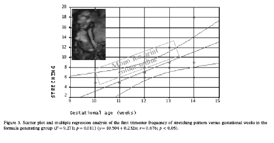
In our study, among eight movement patterns
studied in the first trimester, general
movements followed by isolated hand movements
were the most frequent movement patterns. We
observed a tendency towards an increased
frequency of fetal movement patterns with
increasing gestational age. These findings are
in agreement with the literature. General
movements are the first complex fetal movement
patterns observable by 2D US.
According to Prechtl these are gross
movements, involving the whole body. They can be
recognized from 8 to 9 weeks of pregnancy and
remain present until 16&endash;20 weeks after
birth. Some of the movement patterns could not
be observed through all trimesters. For example,
startleand stretching, which were observed in
the first trimester, disappear with the
progression of pregnancy. As the pregnancy
progresses, the random movements of the
fetalbody, which are the earliest signs of fetal
activity, change into the well-organized
behavioral patterns. Only a few studies are
available on fetal movement patterns during the
second trimester. De Vries and colleagues
studied fetal movements from 20 and from 24
postmenstrual weeks onwards. During the secon
trimester of pregnancy, the incidence of body
movements increased considerably. Kurjak et al.
recently reported the first study that described
the 4D US techniques used for obtaining
longitudina standard parameters of fetal
neurological development in all trimesters of a
normal pregnancy.
They found a tendency towards an increase in
the frequency of fetal movement patterns at the
beginning of the second trimester by 4D US.
However, all types of head movements and hand to
body contact movements indicated a decrease in
frequency from the beginning of the second
trimester to the end of the third trimester. Our
results are similar to this study, as we found a
significant correlation between all head
movements and hand to body contact patterns
during the second and third trimesters except
for head anteflexion, which did not show a
significant change during the second half of
pregnancy. It has also been suggested that there
is a tendency towards decreased frequency of
observed facial expressions and movement
patterns with increasing gestational age.
It has been suggested that the observation
of behavioral quality is a better predictor of
neurological impairment than neurological
examination. In this respect, we are unable to
study the quality of facial movements in
fetuses, because this parameter has not yet been
described. The observation of facial expression
may be of scientific and diagnostic value and
this scientific approach opens an entirely new
field. For example oneof the diagnostic goals of
observing facial expressions is the prenatal
diagnosis of facial paresis. Criteria for the
identification are asymmetrical facial movements
and detection of the movements restricted to
only one side of the face. In one study, the
most frequent fetal and neonatal movements
registered in the third trimester and in the
neonatal period were scowling, eye and mouth
opening, and hand to face, hand to eye, and hand
to head movements.
There was a tendency towards a decrease in
the frequencies of observed facial expressions
(isolated eye blinking, mouthing) and some hand
movement patterns (hand to head, hand to mouth,
hand to face, hand to eye, hand to ear) with
increasing gestational age. Significant trends
in fetal eye movement organization can also be
observed during the second half of pregnancy,
especially during the third trimester
[10,21]. The earliest eye movements
appear at the 16th to 18th weeks of gestation.
At 24 to 26 weeks of gestation, they appear more
frequently. At 36 to 38 weeks of gestation, they
become integrated with other parameters of fetal
activity.
The Zagreb group in another study, evaluated
fetal behavioral patterns in the third trimester
between 30and 33 weeks of gestation in 10
gravidas. They noted that among facial
activities observed by 4D US, simultaneous
eyelid and mouthing movements dominate between
30 and 33 weeks of gestation.
However, another study from the same group
noted that all types of facial expressions
displayed a peak frequency at the end of the
second trimester except isolated eye blinking,
which increased at the beginning of the 24th
week. The fetuses displayed a decreasing or
stagnant incidence of fetal facial expression
from the beginning of the third trimester.
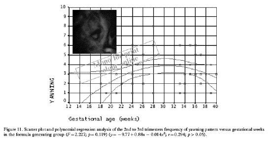
In the present study, while mouthing,
yawning, tongue expulsion, smiling,
sucking, and swallowing expressions displayed a
peak frequency between the 24th and 32nd
gestational weeks similar to the previous
reports, grimacing and eye blinking expressions
displayed peak frequency between the 28th and
36th weeks and after the 32nd week,
respectively. In our study, the fetuses
displayed decreasing or stagnant incidence of
fetal facial expression at the middle of the
third trimester, except for eye blinking, which
showed an increased frequency with increasing
gestational age. This was not in concordance
with the findings of other authors, probably due
to a small sample size in our study and
different study samples in all trimesters of
pregnancy.
The major problem with the study of fetal
behavior is that it is very time-consuming.
Nevertheless, there is no other means of
assessing the function of the central nervous
system in utero, and this is needed for the
understanding of the hidden information in the
neurodevelopmental pathways of the fetal CNS.
Only if normal behavior is understood, is it
possible to identify abnormal behavior before
birth.
Conclusions
Despite all these efforts, it is not yet
clear how we might identify the fetuses with
more specific cerebral damage. Currently there
is no unified neurobehavioral assessment method
for the fetus. The goal of all investigations
should be to gather information that reveals
neural continuity from fetus to newborn. The
availability of quantitative standards might be
important for the experts, to allow an awareness
of normal fetal behavior throughout the whole of
gestation in order to assess the neurological
condition of the fetus.