Abstract : The capacity of
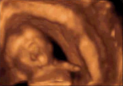
four-dimensional sonography to evaluate complex
facial expressions allows recognition of a
common behavior, yawning. Although there has
been remarkably little interest in yawning in
research and medical practice, even though it is
an everyday phenomenon, we submit an original
interpretation on the basis of knowledge derived
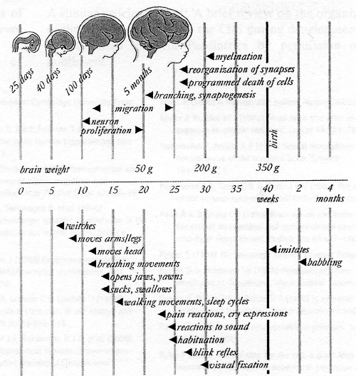
from phylogeny and ontogeny. As a flip-flop
switch, the reciprocal interactions between
sleep- and wake-promoting brain regions allow
the emergence of distinct states of arousal. By
its ontogenical links with REM sleep, yawning
appears as a behavior which procures an arousal
reinforcement through the powerful stretch and
the neuromuscular rewiring induced. Yawning
indicates a harmonious progress in the
development of both the brainstem and the
peripheral neuromuscular function, testifying to
the induction of an ultradian rhythm of
vigilance. The lack of fetal yawn, frequently
associated with lack of swallowing, associated
or not with retrognathia, may be a key to
predict a brainstem's dysfunction after birth.
(en
français)
Résumé:
L'échographie 4D permet
l'évaluation des expressions faciales du
foetus et en particulier de reconnaître un
comportement banal, le bâillement. Bien
qu'il s'agisse d'un comportement
pluri-quotidien, le bâillement a
suscité peu d'intérêt tant
en recherche qu'en pratique médicale.
Phylogenèse et ontogenèse
permettent de proposer une théorie de son
origine. Comme un interrupteur de type "va et
vient", l'alternance d'action des structures
cérébrales stimulant
l'éveil ou le sommeil engendre
l'émergence de différents
états de vigilance. Succédant
à l'hypotonie musculaire du sommeil
paradoxal avec lequel le bâillement
partage des liens ontogénétiques,
l'étirement musculaire puissant qu'il
représente active par
rétro-contrôle les structures du
tronc cérébral impliquées
dans l'éveil. L'existence du
bâillement chez le foetus témoigne
d'un développement harmonieux du tronc
cérébral, de la fonction
neuro-musculaire périphérique et
de l'installation de rythmes ultradiens de la
vigilance. La reconnaissance de l'absence de
bâillements comme de mouvements de
déglutition, associée ou non
à un rétrognatisme prédit
un risque de dysfonctionnement postnatal du
tronc cérébral. (in
english)
Introduction
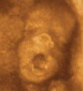
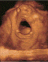
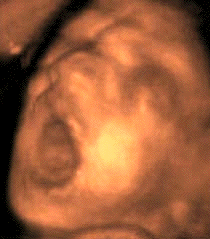
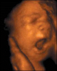
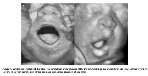
The use of ultrasound examinations during
pregnancy allows a type of fetal behavior,
yawning, to be observed on a daily basis. Few
data have been published in the last 25 years on
yawning, thus prompting researchers to state
"yawning is a universally well known, but poorly
understood" [1] and "a rudimentary
reflex, appears to have at best an obscure
purpose, if any" [2]. Although there has
been remarkably little interest in yawning in
research, even though it is an everyday
phenomenon, we will discuss the meaning of this
behavior and how its characterization can
enhance ultrasound investigation. As a foreword,
it should be noted that human research on
prenatal programming of behavior is
intrinsically correlational, never
manipulatively experimental, and frequently
based upon homologies with other
vertebrates.
A popular saying states that "the organ
generates the function". However, it is known in
embryology that body movement in a fetus is
required for maturation of the motor function
and is involved in the development of other
organs such as the lung. On the other hand, body
movement indicates a harmonious progress in the
development of both the central motor system and
the peripheral neuromuscular function
[3].
All the movements that a newborn is able to
produce originate during the fetal life and are
performed throughout the life of the individual.
Behaviour observed in utero, including
breathing, yawning, and others, serves as a
continuum to the activity shown in a newborn
infant and undergoes a neuromuscular rewiring
[4]. The onset and developmental course
of fetal motility have been studied since the
introduction of ultrasound technology. Four
dimensional ultrasonography (4D-US) provides a
tool for movement observations not only for
their differentiation but also to categorize
specific patterns of behavior. The evaluation of
facial expressions was previously impossible
using real-time two-dimensional ultrasonography.
The capacity of 4D-US to evaluate complex facial
expressions helps to identify a common behavior,
yawning [5,6].
What yawning is not
Provine and Tate [7] found no
support for the popular hypothesis that yawning
is a response to elevated CO2 or depressed 02
levels in the blood. Subjects breathing pure
oxygen did not show a decreased amount of
yawning. Studies on fetal yawning (like a fish
yawn in water) in amniotic fluid do not make any
association between oxygenation capacity and
yawning. Yawning occurs frequently when a fish
is interrupted in feeding, for example by the
approach of a female and before courtship is
initiated. This behavior does not need any
particular level of oxygenation.
Atelectasis, the collapse of alveoli,
results from alveolar hypoventilation of air.
The normal respiratory pattern of spontaneously
breathing adults includes periodic sighs or deep
breaths (also a type of yawning) that prevent
atelectasis by producing alveolar surfactants.
But yawning confers no protection against
atelectasis in a fluid-filled lung of a fetus
which produces surfactant at the time of first
breath after birth [8].
How to recognize a yawn
A yawn is a paroxystic cycle characterized
by a standard cascade of movements over a 5-10 s
period:
ample, slow, and very deep inspiration,
mouth wide open (Figure 1); in human adults, the
expansion of the pharynx can quadruple its
at-rest diameter, while the larynx opens up with
maximal abduction of the vocal cords
a brief interruption, the acme state, often
with accompanying limb and neck stretching;
a rapid passive expiration [9].
4D-US helps to identify this typical
development: the fetal mouth, previously closed,
opens widely for 4-6 s with simultaneous
retraction of the tongue, followed by rapid
closure, and mostly combined with retroflexion
of the head and sometimes elevation of the arms
(pandiculation) (Figure 2). This harmonious
sequence is markedly different to a brief
swallowing episode [10]. Using a
color Doppler technique, it is possible to
observe the flow of amniotic fluid through the
fetal mouth, oropharynx, pharynx, and trachea to
the lungs. This movement pattern is
non-repetitive in the fetus, in contrast with
that in adults [11].
Yawning is not just a matter of opening
one's mouth, but a generalized stretching of
muscles, those of the respiratory tract
(diaphragm, intercostal), face, and neck (Figure
3). This association of complex and synergic
movements is a very stereotypical behavior that
can be classified as a reflex due to its
involuntary occurrence. The reflex arc is
thought to be in the hypothalamus, the reticular
system in the brainstem, and it involves the
respiratory neurons in the medulla, the motor
nuclei of die 5th, 7di, 9th, 10th, and 12th
cranial nerves, the phrenic nerves (Cl-C4), and
the motor supply to the intercostal muscles.
Also, it can be inferred that yawning is a part
of the generalized stretch that generally
accompanies the yawn [9].
Embryology and mechanisms
In 1973, T. Dobzhansky remarked: "nothing in
biology makes sense except in the light of
evolution" [12], and Ernst Haeckel
(1834-1919) stated "ontogenesis is a brief and
rapid recapitulation of phylogenesis, determined
by the physiological functions of heredity
(generation) and adaptation (maintenance)"
[13]. The exactitude of these quotations
is illustrated by yawning. Indeed, the
ultrasound investigation specifies is
ontogenesis precociousness between 12 and 15
weeks of gestation (Figure 4) [4].
Indeed, yawning is also a phylogenetically old,
stereotypical event that occurs in reptiles,
fish, birds, and mammals. Its survival without
evolutionary variations postulates a particular
importance in terms of developmental needs
[9]. The strong muscular contraction
that signifies a yawn is metabolically
expensive. If we accord with the terms of
Darwin's evolutionary propositions, the costs of
brain activity must be outweighed by the
advantages gained in terms of developmental
fitness. Thus, a structural hypothesis suggests
activation in the synthesis of neurotrophins,
which lead to a cascade of both new synapse
formation or recruitment, and activation through
the diencephalon, brainstem, and spinal cord.
The phenomenon of activity-dependent development
has been clearly shown to be one mechanism by
which early sensory or motor experience can
affect the course of neural development.
Activity-dependent development may be a
ubiquitous process in brain maturation in which
activity in one brain region can influence the
developmental course of other regions
[14]. The ability to initiate motor
behavior generated centrally and linked to
arousal and respiratory function is a property
of the brainstem reticular formation, which has
been remarkably conserved during the phylogeny
of vertebrates including agnathans, fishes,
amphibians, reptiles, and birds. Therefore,
conservative developmental mechanisms
orchestrating the organogenesis of the brainstem
in all vertebrates are probably crucial for
arousal and breathing [15].
A wealth of data have accumulated on genes
that are expressed in the embryo and govern the
hindbrain segmentation. Hox homeobox genes form
four conserved clusters encoding transcription
factors that orchestrate ontogenesis along the
rostrocaudal axis of the body, including
hindbrain segmentation and limb formation
[16]. This might explain craniofacial
congenital developmental abnormalities that
ultrasound investigation helps to reveal; hence,
"the face predicts the brain".
The facial bone structure and the brain
differentiate from a common embryonic structure,
the ectoblast. The cephalic pole comprises an
original embryological encephalo-facial and
encephalocervical segmentation with a strict
topographical correspondence: the naso-frontal
and premaxillary structures are joined to the
forebrain; and the maxillo-inandibular and
anterior cervical structures are joined to the
brainstem and its nerves. At the beginning of
the third month, the embryo becomes a fetus with
the occurrence of the first oral and pharyngal
motor sequences under the control of the
neurological development of the brainstem,
development of the suction-deglutition, and
yawning activity (Figure 4). Therefore, suction
and yawning have the same embryological origin,
thus demonstrating the importance of the
brainstem in the neurophysiological development
of the oropharyngeal activity coordinated with
the respiratory, cardiac, and digestive
regulations which have the same neuroanatomical
localization. Its occurrence markes the
developmental stage when the brainstem is
already individualized and the pituitary gland
has become functional, whereas the extension of
the temporal and frontal neocortex takes up to
22-24 weeks to reach completion
[17,18].
Movements of the tongue or jaws assit the
development of the palate by promoting the
horizontal elevation of vertically orientated
palatal shelves (Figure 3). Activity of the neck
and tongue muscles is always accompanied by
mouth-tongue movement [19]. The
relationship between the neural network of
mouth-tongue movement and respiratory activities
is not perfectly understood. It seems that
information about central respiratory and
locomotor rhythms that is necessary for
cerebellar control of the coordination between
respiration and locomotion converges at the
level of the lateral reticular nucleus
[20].
Yawning is under the control of several
neurotransmitters and neuropeptides at the
central level. The paraventricular nucleus of
the hypothalamus is the hypothalamic center,
which adapts and coordinates hormonal and
autonomic responses for the appropriate
behavior, and controls yawning. Oxytocinergic
neurons are stimulated by dopamine, excitatory
amino acids, acetylcholine, serotonin, nitric
oxide, and adrenocorticotropic hormone-related
peptides (all implicated in arousal), while
opioid peptides inhibit this behavior. They
project to the hippocampus, and the reticular
formation of the brainstream, which play a key
role in the expression of this behavioral event.
Other neurotransmitters, i.e., floradrenaline
and neuropeptides, hypocretin and sexual
hormones, influence this behavioral response
[21].
Why yawning shares a link with
arousal
The phylogenetic appearance of sleep
proposes that the nocturnal resting in
poikilotherms most probably manifests in mammals
as a form of rapid eye movement (REM) sleep or
paradoxical sleep, which is characterized by
peripheral muscular atonia originating in the
dorsal part of the brainstem, rostral to the
pons [22].
Based on numerous studies of fetuses and
infants in a variety of mammalian species, it is
widely believed that the earliest form of sleep
is properly characterized as active sleep, that
is an immature form of REM sleep and
preponderant at birth. Accordingly, it is
thought that quiet sleep, an immature form of
slow-wave sleep (SWS), emerges as REM sleep's
predominance diminishes during ontogeny
[23].
In the early intra-uterine life, a diffuse
collection of phasic and cyclic motor events
occur that gradually coalesce. For the fetus,
sleep and wakefulness are reliably
characterized, respectively, by periods of
myoclonic twitching expressed against a
background of muscle atonia and high-amplitude
behaviors (e.g., locomotion or
stretching-yawning) expressed against a
background of high muscle tone. Movements of the
limbs, such as stretching, yawning, and kicking,
are typically considered to indicate periods of
wakefulness. Periods of twitching are almost
always followed by the abrupt onset of
high-amplitude awake behaviors, thus completing
the cycle. Although myoclomc twitching during
active sleep in infants is more prevalent and
more intense than that seen during REM sleep in
adults, its similarities to the adult behavior
and its linkage to periods of atonia suggest
developmental continuity between the infant and
adult sleep states. The maturation of the
central nervous system, based on myelinization,
starts in the spinal cord and then proceeds to
the brainstem and forebrain. Thus, paradoxical
sleep mechanisms located in the brainstem are
the first to mature and the only ones to
function. Then, the slow-wave sleep and waking
structures become mature. Namely, the widespread
control of neuronal activity exerted by specific
REM sleep processes help to direct brain
maturation through activity-dependent
developmental mechanisms. It may be inferred
that REM sleep (and possibly yawning) directs
the course of brain maturation in early life
through the control of neural activity
[24].
Behavioral pattern continuity from prenatal
to postnatal life shows a strict parallelism
between the ontogeny of REM sleep and yawning
(Figures 5 and 6) [6]. Basically, REM
sleep in the human declines from 50% of total
sleep time (8 h) and a frequency of 30-50 yawns
per day, in the newborn, to 15% of total sleep
time (1 h) and less than 20 yawns per day, in
the adult. This decrease takes place mainly
between birth and the end of puberty
[25].
The emergence of distinct states is followed
by dramatic changes in the amounts, duration,
and cydicity. An ultradian rhythm may be graded;
in a period from 50 to 60 minutes appears an
ALTernation of moment characterized by motor
activity and by rest, as in newborns. Each
period of rest switches over a period of
activity by a yawn. Thus a periodicity of one or
two yawns per hour can be noticed [4].
Yawning appears 2 weeks before any discernible
sleep-wake states, and its expression gradually
becomes linked. No changes in the incidence of
yawns between 20 and 36 weeks of gestational age
have been observed by Roodenburg and colleagues
[26] in the fetus. In full-term infants,
yawns are frequentely observed on the first day
of life. The embryo and fetus are exposed to 24
h periodicity with the mother's parameters of
the circadian cycle, which may play a role in
the normal development of the fetal pacemaker.
There are no data available for how a fetal's
yawn links up fetal rhythm with maternal
rhythm.
Saper and coworkers [27] propose a
model for reciprocal interactions between sleep-
and wakepromoting brain regions, which produces
a flip-flop switch. This model could explain the
rapid transitions from awaking in sleep and from
REM sleep to waking. From a survival point of
view, it is necessary to ensure that there is a
period of sleep for body repair to take place
but also for the individual to have the capacity
to flee a predator (arousal). The transition is
controlled by integrative autonomic structures
that encompass regulated changes occurring in
anticipation of the event. Yawning (a stretch
syndrome) can be seen as a behavior, a testifier
of this switch/transition, like a reinforcement
of muscle tone. Waking is controlled by some
four different and redundant circuits mainly
located in the reticular formation of the pons
(adrenergic), the peduncle (dopantinergic), the
hypothalamus (histaminergic), and the Meynert
basifrontal region (cholinergic). The permissive
networks controlling waking must be tonically
reinforced by the hypocretin's system
originating from the lateral hypothalamus. Next,
neuronal activation of the ventrolateral
preoptic nucleus (\TLPO) is correlated with the
amount of sleep. The powerful muscular
contraction caused by yawning releases arousal
by activation of the reticular-formation (locus
coeruleus) to which the cranial nerves send
retroprojections. At awakening, the yawning and
stretching reverse the muscular atonia which
characterize REM sleep. On the other hand, when
the pressure to sleep increases, it is thought
that the firing of the GABA and galanin VLPO's
neurons reduce the muscular tone of
antigravitational muscles, notably those of the
neck and masseters. Thus, yawning seems to be
averse to this pressure. F. Giganti and
coworkers observed yawning in premature infants
in all behavioral states, except during quiet
sleep, and viewed it as a transitional state,
suggesting a spreading activation of facial
motor patterning. Thus, yawning may be seen as a
nervous reflex loop which occurs as arousal
reinforcement [28-30].
Yawning or not: A pathology?
Yawning occurs with a regular recurrence,
about once or twice per hour. When a yawn is
observed during a 4D-US examination, it is
obviously by chance or after a very long
investigation (Figure 6). Yawning appears
preferentially after a period of rest and
indicates awakening. If normal swallowing is
seen (much more frequent), the search for
yawning glossoptosis. Several arguments favor an
embryonic origin consisting of an anomaly in the
caudal hind-brain development. Feeding disorders
are the most important functional symptom.
Testimonies from mothers seem to agree with the
lack of yawning at birth and a paralell progress
during the first year of life in swallowing and
yawning. Also, Pierre Robin syndrome can be seen
as prenatal brainstem dysfunction responsible
for the oro-facaial maldevelopment which can be
diagnosed at 23 weeks' gestation during a 4D-US
examination [33,34].
Any syndrome (primary bilateral or
unilateral growth anomalies) associated or not
with temporo/ mandibular joint ankylosis,
aglossia/microglossia: Francheschetti syndrome,
Goldenhar syndrome, and Richner-Hanhart
syndrome.
The Moebius syndrome is characterized by
congenital facial diplegia and bilateral
abducens nerve palsies by degenerative and
involved nuclei of the VI, VII, and XII nerves.
Simultaneous occurrence of limb malformations
with cranial nerve dysfunction suggests a
disruption of normal morphogenesis during a
critical period in the development of the
embryonic brainstem, most likely from 4 to 7
weeks of gestation. Instances of bilateral
paresis of the soft palate and scattered
instances of dysphagia (some of which resolve in
infancy) have been reported. An inability to
close the mouth is the norm [35].
Watershed infarcts in the fetal and neonatal
brainstem are clinically expressed as multiple
cranial neuropathies, failure of central
respiratory drive, dysphagia [36].
Goldenhar Syndrome includes malformations
primarily involving the jaw, mouth, and ears,
and, in most cases, affect one side of the body.
This represents defects in the embryonic first
and second brachial arches, the first pharyngeal
pouch and brachial cleft, and the primordia of
the temporal bone [37].
Joubert syndrome is a rare, genetic disorder
characterized by an absence or underdevelopment
of the cerebellar vermis and a malformed
brainstem. The most common features include
ataxia, an abnormal breathing pattern, sleep
apnea, abnormal eye and tongue movements, and
hypotonia.
It is possible to complete this catalog by
referring congenital trismus, Crisponi syndrome,
SniveWiedemann syndrome.
Conclusion
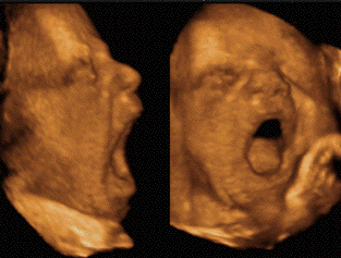
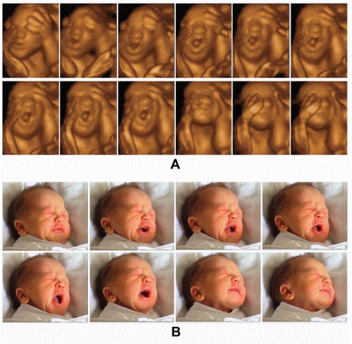
With significant advances in image quality,
resolution of ultrasound, and now 3D and 4D
technology, the use of ultrasound examination
during pregnancy is a step forward from
anatomical examination to functional evaluation.
Recognition of fetal yawning aids to testify of
the harmonious progress of brainstem maturation
and to understand the neural underpinnings of
sleep and arousal systems. An abnormality yawn's
occurrence fosters an intensive research of
anemic fetuses (frequency amplified) or
brainstem dysfunction with or without mandibular
hypoplasia (frequency sparse or null). We hope
and expect that upcoming researches will
complete the data currently available.
5. Kurjak
A, Stanojevic M, Azumendi G et al. The
potential of four dimensional (4D)
ultrasonography in the assessment of fetal
awareness. J Perint Med. 2005;33:46-53.
6.Hata
T, K Kanemshi, et al. Real-time 3-D
sonographic observation of fetal facial
expression J Obstet Gynaecol Res 2005; 31; 4;
337-340
11. Masuzaki H,
Masuzaki M. Color Doppler imaging of fetal
yawning. Ultrasound Obstet Gynecol.
1996;8(5):355-356.
12. Dobzhansky T. Nothing in biology makes
sense except in the light of evolution. The
American Biology Teacher 1973;35:125-129.
13. von
Haeckel E. Anthropogenie oder,
Entwickelungsgeschichte des menschen, Keimes-
und stammesgeschichte. Leipzig : W. Engelmann
ed. 1877: 770p.
14. Briscoe J, Wilkinson DG. Establishing
neuronal circuitry: hox genes make the
connection. Genes Dev.
2004;18(14):1643-1648.
15. Graham A. The development and evolution
of the pharyngeal arches. J Anat.
2001;199:133-141.
16. Köntges G, Lumsden A.
Rhombencephalic neural crest segmentation is
preserved throughout craniofacial ontogeny.
Development. 1996;122:3229-3242.
17. Jacob J, Gutrhie S. Facial visceromotor
neurons display specific rhombomere origin and
axon pathfinding behavior. J Neuosci.
2000;20:7664-7671.
18. Santagati F, Rijli F. Cranial neural
crest and the building of the vertebrate head.
Nature Rev Neurosci. 2003;4: 806-818.
19. Wragg LE, Smith JA, Borden CS. Myoneural
maturation and function of the fetal rat tongue
at the time of secondary plate closure. Arch
Oral Biol. 1972;17:673-682.
20. Ezure
K, Tanaka I. Convergence of central
respiratory and locomotor rhythms onto single
neurons of the lateral reticular nucleus. Exp
Brain Res. 1997;113:230-242.
22. Argiolas
A, Melis MR. The neuropharmacology of
yawning. Eur J Pharmacol. 1998;343(1):1-16.
21. Siegel JM. Sleep phylogeny : clues to
the evolution and function of sleep. In Luppi PH
ed. Sleep : circuits and functions. CRC Press
2005;9:163-176.
23. Valatx JL. The ontogeny and physiology
confirms the dual nature of sleep states. Arch
Ital Biol. 2004;142(4):569-80.
24. Blumberg MS, Luca DE. A developmental
and component analysis of active sleep. Develop
Psychobiol.1996;29(1):1-22.
25. Kobayashi T, Good C, Mamiya K, et al.
Develppment of REM sleep drive and clinicals
implications. J Appl Physiol.
2004;96:735-746.
26. Roodenburg
PJ, Wladimiroff JW, van Es A et al.
Classification and quantitative aspects of fetal
movements during the second half of normal
pregnancy. Early Hum Develop.
1991;25:19-35.
27. Saper
CB, Chou TC, Scammell TE. The sleep switch:
hypothalamic control of sleep and wakefulness.
Trends Neurosci. 2001;24(12):726-31.
29. Pace-Schott EF, Hobson A. The
neurobiology of sleep: genetics, cellular
physiology and subcortcal networks. Nature Rev
Neurosci. 2002;3(8):591-605.
30. Baenninger
R. On yawning and its f Baenninger R. On
yawning and its functions. Psychonomic Bul Rev.
1997;4(2):198-207.
31. Petrikovsky
BM, Kaplan GP, Holsten N. Fetal yawning
activity in normal and high-risk fetuses: a
preliminary observation. Ultrasound Obstet
Gynecol. 1999;13:127-130.
32. American Thoracic Society. Idiopathic
congenital central hypoventilation syndrome:
diagnosis and management. Am J Respir Crit Care
Med 1999;160:368-373.
33. Abadie V, Morisseau-Durand M. Brainstem
dysfunction: a possible neuroembryological
pathogenesis of isolated Pierre Robin sequence.
Eur J Pediatr. 2002;161(5):275-280.
34. Matsumato M, Yanagihara T et al.
Antenatal three-dimensional sonographic features
of Pierre Robin Syndrome. Case report. Gyncol
Obstet Invest. 2001;51(2):141-142.
35. May M, Schaitkin B, Shapiro A: Facial
nerve disorders in newborns and children. In The
Facial Nerve. Thieme Medical Publishers 2000;
2nd edition:339-365.
36. Sarnat HB. Watershed infarcts in the
fetal and neonatal brainstem: an aetiology of
central hypoventilation, dysphagia, micrognatia.
Europ J Paed Neurol. 2004;8(2):71-87.
37. Volpe P, Gentile M. Three dimensional
diagnosis of Goldenhar syndrome. Ultrasound
Obstet Gyncol. 2004;24(7):798-800.
Hata T,
K Kanemshi, et al Real-time 3-D sonographic
observation of fetal facial expression
J Obstet Gynaecol Res 2005; 31;
4; 337-340
Holditch-Davis
D et al Development
of behaviors in preterm infants: relation to
sleeping and waking Nursing
Research 2003; 52; 5;
307-317
Kurjak
A, Azumendi G, et al Fetal hand movements
and facial expression in normal pregnancy
studied by four-dimensional sonography
J.Perinat Med2003; 31;
496-508
Kurjak
A, Azumendi G, et al The potential of 4D
ultrasonography in the assessment of fetal
awareness J Perinat Med 2005;
33; 46-53
Kurjak
A, M Stanojevic et al Behavioral pattern
continuity from prenatal
to postnatal life a study
by four dimensional (4D) ultrasonography
J Perinat Med2004; 32;
346-353
Masuzaki H
Color Dopplerimaging of foetal yawning
Ultrasound in obstetric &
gynecolgy 1996; 8; 5; 355-6
Peirano
P, Algarin C, Uauy R Sleep-wake states and
their regulatory mechanisms throughout early
human development J Pediat 2003;
143; 4S; S70-79
Petrikovsky BM,
Kaplan GP, Pestrak H The application of
color Doppler technology to the study of fetal
swallowing Obstet Gynecol 1995;
86; 605-8
Petrikovsky
B, Kaplan G, Holsten N Fetal yawning
activity in normal and high-risk fetuses: a
preliminary observation.
Ultrasound Obstet Gynecol 1999;
13; 2; 127-130
Roodenburg
PJ et al Classification and quantitative
aspects of fetal movements during the second
half of normal pregnancy Early
Human Development 1991; 25; 19-35