Three-dimensional ultrasound has been
available for more than ten years. During its
continuous improvement and development, several
different kinds of modes have been created. They
include multiplanar imaging, volume rendering,
surface rendering, three-dimensional color
Doppler, three-dimensional volumetry, cine-loop
animation, post-processing and cutting. However,
the three-dimensional image freezes the object
and therefore does not provide information on
movements or any information about the dynamic
changes of the object of interest. A technique
was needed that would enable three-dimensional
imaging to be preformed in a real-time mode.
This technique can be called live
three-dimensional ultrasound (3D-US) or
four-dimensional ultrasound (4D-US), as coined
by a manufacturer, because time becomes a
parameter within the three-dimensional imaging
sequence. Human eyes are known to be able to
differentiate between images up to a frequency
of about 12 images per second, consequently
production of an appropriate frame rate with
specially designed probes and a fast computer
rendering device is required.
At the moment 4D-US scanning is not
real-time and available machines can reach up to
about 20 images per second, depending on volume
size, resolution and the mechanics of the probe.
Nevertheless, even at these relatively slow
frame rates the ability to study fetal activity
is strikingly good.This new diagnostic tool is
enabling the continuous monitoring of the fetal
face and other surface features of the fetus
such as fetal extremities, thus opening up
exciting new possibilities for the study of the
relatively unexplored area of fetal behavior as
a possible measure of neurological maturation.
Four-dimensional sonography (4D-US) provides a
new tool for observation of movement
differentiation. The developmental pattern of
hand movement over the first phase as seen by
4D-US has been described elsewhere.However, the
differentiation of hand movement over the second
phase has not been observed by 4D-US.This topic
was thus the focus of interest in the current
study.
In the early second trimester 4D-US provides
simultaneous visualization of all four
extremities and enables confident recognition of
isolated arm movements and their direction.
Because of the limitations of 2D-US only five
types of isolated hand movements can be
described. They include: hand to head, hand to
trunk, hand to foot, hand to fluid and hand to
the uterine wall. If one performs 4D-US hand to
head movement can be differentiated into seven
subgroups: hand to head, hand to mouth, hand
near mouth, hand to face, hand near face, hand
to eye and hand to ear. We determined the
incidence of each subtype of isolated hand to
head movements between 13 and 16 weeks of
gestation. Simultaneous imaging of complex
facial movements and the evaluation of facial
expression was impossible using real time 2D-US.
On another hand, 3D-US provides images with
recognizable facial expression, although it
remains impossible to determine the duration of
facial activity. 4D-US integrates the advantages
of the spatial imaging of the fetal face with
the addition of time.This novel technology
therefore allows the appearance and duration of
each facial movement and expression to be
determined and measured.
[...]
yawning Ð slow and prolonged wide
opening of the jaws followed by quick closure
with simultaneous retroflexion of the head and
sometimes elevation of the arms of exoration;
tongue expulsion Ð facial activity
characterized.
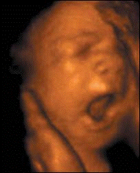
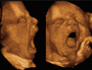
Surface rendering by 3D-US demonstrates fetal
yawning. Note clear demonstration of the
contours of the fetal lips, and the oro-facial
muscles underneath, which cause this
movement.The surface of the tongue is also
visualized. The entire dynamics of the
oro-facial region, however, can only be observed
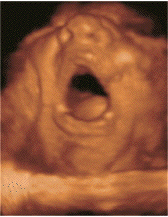
with 4D-US.Figure 6. On this image sequence, the
same movement pattern is shown by 4D-US.Yawning
could be observed as a movement pattern
identical to that seen in infants, children and
adults. On a figure sequence one can see head
movements (deflexion and rotation) associated
with slow opening, prolonged wide opening of the
jaws, followed by quick closure with a
simultaneous retroflexion of the head.
On this image sequence, the same movement
pattern is shown by 4D-US.Yawning could be
observed as a movement pattern identical to that
seen in infants, children and adults. On a
figure sequence one can see head movements
(deflexion and rotation) associated with slow
opening, prolonged wide opening of the jaws,
followed by quick closure with a simultaneous
retroflexion of the head.