Anatomiste de talent, Bartolomeo
Eustachi a fait progresser cette science dans la
seconde moitié du XVIe siècle. Outre la
fameuse trompe d'Eustache, il a
révélé l'existence de la valvule qui
porte son nom, des surrénales, du canal
thoracique.
Le grand oeuvre de Bartolomeo Eustachi devait
être un traité d'anatomie « De
dissensionibus ac controvesiis anatomicis ». Il
devait comporter 47 planches anatomiques,
dessinées avec l'aide de Pier Matteo Pini,
richement détaillées et
légendées. Seulement 8 planches furent
publiées de son vivant. Les 39 autres, perdues,
ont été longtemps recherchées. Elles
ont été retrouvées 162 ans plus tard
chez un descendant de Pier Matteo Pini. Publiées
en 1714 sous le titre « Tabulae anatomicae
Bartolomaei Eustachi quas a tenebris tandem vindicatas
» (illustrations anatomiques de Bartolomeo Eustachi
sauvées de l'obscurité), elles font de leur
auteur, avecVésale, l'un des pères de
l'anatomie moderne.
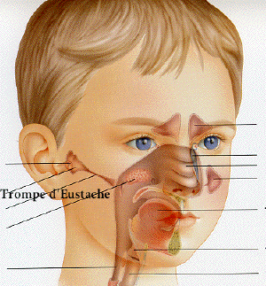
On a discuté de l'anatomie et de la
physiologie de la trompe d'Eustache. Les
observations salpingoscopiques ont prouvé
que le tube Ostium pharyngeum s'ouvre largement
à chaque bâillement; une grande
quantité d'air est donc poussée
dans la cavité tympanale. Ce
phénomène est marqué par
une dureté d'oreille considérable,
des bruits graves, de l'autophonie causée
par l'augmentation de la pression d'air vers la
fenêtre et la membrane tympanale dont les
mouvements pourraient être
enregistrés à l'aide du
manomètre à oreille de
Politzer.
Le diagramme des mouvements en question est
très différent quand nous
bâillons par une profonde aspiration par
le nez et une courte expiration par la
bouche.
La perte de son du muscle
ptérygoïde interne dans la
subarthrose, dysarthrose et dans les
mâchoires édentées,
influence de la même façon les
muscles tensor veli palatini et tensor tympani,
causant une diminution de l'aération de
l'oreille moyenne avec une déperdition de
l'ouïe consécutive, ca.15-20db.
Quant à la disparition rapide de la
surpression dans la cavité tympanale
pendant le bâillement juste à la
fin d'une expiration prolongée, la
contraction du muscle tensor tympani est
responsable de ce phénomène. Le
relachement du dit muscle dans le
bâillement comme accompagnement de la
contraction et du relâchement du muscle
orbicularis oculi présente une
diiminution relativement lente jusqu'à ce
que le muscle soit détendu
complètement. L'enregistrenient du
changement de tension du muscle tensor tympani
pourrait être essayé en mesurant
les impédances acoustiques de l'appareil
de transmission après l'irritation de
l'oreille donnée par des sons forts ou
intermittents, comme l'ont prouvé 0.
Metz, Perlmann et Casse.
Yawning is a somatical reflex serving the
purpose of augmenting the tonus of the limbs and
trunk muscles no less than the negative pressure
in the mediastinum, which promotes the outflow
of venous blood from the filled-up veins of the
distant parts of the body. It also must be
considered as an expression of the feeling of
exhaustion and weariness. An organ taking an
active part in yawning is the Ear. It is a
common knowledge that as long as one yawns
nothing can be heard, and when the yawning
reflex is finished the hearing returns quickly.
The explanation of this phenomenon must be
sought in the relation between the mechanism
producing opening of the Eustachius tube to the
aeration of the tympanal cavity as well as the
contraction of the tensor tympani and stapedius
muscles in yawning. The Eustachius tube forms a
funnel-like canal leading from, the naso-pharynx
to the midde Ear an extent of ca. 35 mm in the
adult. The diagonal course of its half inch
short bony part and the angular descent of ca.
40 degrees from the horizontale i. e. the base
of the skull of its 3 lines longer
cartilagineous part forms some complicating
topography of the tube in question.
The cartilagineous portion when seen in
cross section looks like a sheperd's crook
(Hirtenstabkrummung of the German authors). The
short lateral cartilage serve as a fine of
insertion for the tensor veli palitini (m.
spheno-salpingostaphylinus), arising from fossa
scaphoidea of the sphenoid bone. Levator veli
palatini, forming the bottom of the tube starts
from the inferior apical part of the pyramidbone
and then is called
petrosalpingo-staphylinus.
Urbantschitsch has compared the lumen of the
tube to the flat double pens the narrowing of
which (istmus) shows many variations. The
collapsed soft tissue (pars membranacea) of the
pharyngeal end of the Eustachius tube is
believed by Armstrong to close its lumen when
the mentioned muscles are in rest (flutter valve
mechanism). On swallowing or yawning the tensor
veli palatini opens booldike the walls of the
Eustachius tube while the bulge of the levator
veli palatini supports the separation of its
walls. The nerves supplying tensor tympani,
levator, tensor-veli palatini and pterygoideus
intern. muscles derives from the anterior
part of the 3d branch of trigeminus (portio
minor trigemini or nervus masticatorius).
Therefore the synchronisation of their
innervation inspite of many differences consists
in their motion. The loss of tone of the
pterygoideus. int. muscle, which appear often in
cases of malformation of the temporo-mandibular
joint as s.c. subarthros and dysarthros in
edentelous jaws, influences in the same way the
tensor veli palatini and tensor tympani muscles.
Thesefunctiona] changes are manifested by
the diminished aeration of the middle Ear on the
given side with noises, tinnitus, loss of
hearing for whisper, speech and tunning forks
under 4000 Hz. The acrylitic splint fort the
purpose of completing the edentelous bite
applied by Ronkin in these cases, had increased
the tonus of pterygoideus int. muscle through
the head noises and tinnitus has markedly
decreased, rough which the 15 db. improvement of
hearing was reached.
Change of tonus of levator veli palatini m.
has been described by Gyergay with susbsequent
wide opening of the Eustachius tube during
swallowing, which causes a greater amount of air
to be driven into the middle Ear than is
necessary for compensation of the negative air
pressure there.
Salpingoscopie observations proove that the
ostium pharyngeum tubae opens widely in
yawning or swallowing by retraction of
its anterior wall. Repeated swallowing reveals
that the closed ostium does not open every time.
In yawning however it opens every time to such
an extent that a great amount of air is driven
into the tympanal cavity. This phenomenon is
marked by a considerable hardness of hearing low
noises, autophony caused by increased air
pressure towards the fenestra (rotundum and
ovale) as a compliant part of the rigid wall of
the tympanal cavity. This fact elicits changes
in the peri-endolymph pressure, lasting as long
as the superflous amount of air is discharged by
the action of the tympanic muscles : tensor
tympani, stapedius. In order to prove the
influence of yawning on the variations of air
pressure in the tympanal cavity as well as
the action of the tympanal muscles in yawning I
have tried to register the movements od the
tympanal membrane with the help of an Ear
manometer resembling the one used by
Politzer.
The level of alcohol-fuschin sol. placed in
the lumen of the U shaped glass tube of this
manometer enabled us to read the variations of
air pressure in the external auditory canal as a
consequence of movements of the tympanal
membrane in yawning With help of the background
mililmeter scale the diagram could be
established, which illustrates approximately the
problem in question. When we yawn as a deep
inspirium, through the nose and a short expirium
by the mouth, the diagram of the tympanal
membrane movements shows a lengthened contours
from the upper to the lower end. It is evident
that the ostium of the Eustachius tube is
principally closed and the resistance of it
closure could be proved by the imethod of
Zöllner (overpressure in the nasopharynx
and simultaneously observing the movements of
the Eardrum) and the method of v. Dishoeck by
using of pneumophone. The said autbor has
prooved that the pressure 30-60 cm of water in
Valsalva or Politzer test, is able to overcome
this resistance, on the contrary the
overpressure in the tympanic cavity caused by
swelling or catarrhal inflammation of the
tympanal mucosa pass easier through the
tightness of the obstructed tube by using 12 cm
of water only. The air caught in the typanum
will be resorbed til itequals the pressure of
the blood-gasses. Partial pressure of the
capillary bloodgasses are : Oxygen.... 90 cm of
water;... 800 cm w.; CO2 60 cm; total 950 cm w .
Atmospheric pressure on the outside of the
tympanal membrane amounts 1000 cm. of water; the
differential negative pressure in the tympana
cavity is approximatively 50 cm of water which
could be easily proved by using of the mentioned
pneumophone and the hearing disturbances
establislied mainly for the frequencies under
4000 Hz. as 15-20 db. loss. The high tones are
improved in negative as well as in positive
pressure differences in the tympanic cavity. It
is striking that the overpressure in the
tympanuin in yawning in normal individuals
causing a considerable hardness of hearing
disappears quickly just a the end of the
prolonged expirium in this phenomenon.
The explanation of this fact must be sought
in the contraction of the tensor tynipani muscle
which facilitated the expulsion of the
superflous amount of air from the tympanal
cavity. The start of the contraction in question
is synchronous with levator and tensor veli
palatini, but the relaxation of the tympanal
muscle is not contemporary with the said
muscles. Perlmann, Casse and Metz have proved
that the relaxation of the afore mentioned
muscle after irritation of the given Ear with
strong or interrupted tones, lasts relatively
long until this muscle unbends completely.
Likewise, the spontaneous contraction of tensor
tympani m. in yawning as a concomitant to that
of orbicularis, oculi m. presents a much more
slow decrease of the contraction curve than when
it is produced acoustically. This could be
tested by the registration of the change in the
tension of the tympanal membrane rneasuring
acoustic impedances of the transmission
apparatus, which is to be compared with
avariable acoustic impedance standard and the
Ear of the examiner as it was described by
0.Metz ( in Acta Oto-Laryngol., Supplementum 63,
and vol. XXXIX, f. 5). This apparatus consists,
of a tube ends of which are adapted to the Ear
of the subject and the opposite one to the
examiners. The telephone-membrane placed in the
mesial part of this tube sounds to either side.
By means of a Y shaped glass-tube this acoustic
bridge is connected with audio-oscillator,
piezo-electric microphone, amplifier and the
string oscillo-graph. The opposite Ear of the
subject is attached to the audiometer-telephone
as a source of acoustic stimulus, producing, the
change in the tension of the tympanic membrane.
According to Lorente de No this unilateral
acoustic stimulus elicit bilateral contraction
of tensor tympani m. resembling a pupillary
reflex.
The intensity of tone from audio-oscillator
at the detector (piezo-electric
microphone-amplifier and oscillograph) gives
approximately a notion of the change in
impedance of this acoustical transmission,
caused by the contraction of the tympanal mscle.
In order to test changes in tension of the
tympanic membrane related to the contraction of
the tensor tympani muscle in yawning an
audiometer telephone could be eliminated from
the above described apparature used by 0. Metz
and replaced by a rubber tube only, joining this
Eaar with the main tube. By using such a
simplified apparatus oscillogiaphic records of
changes in the impedance of the tympanic
membrane could be given, produced by contraction
of the tensor tympani muscles in yawning. This
sound curve shows a slow rising, and ceases just
as slowly as the analogous one produced
acoustically by 0. Metz.
This oscillographical record-cuve of the
impedance in question confirms the supposition,
that the prolonged contraction and relaxation of
the tensor tympani muscle, lasting for a long
time just after the ceasing of expirium phase in
contributes in a great measure to the expulsion
of the superflous amount of air from the
tympanal cavity by which a « quick Ear
» is promptly restored. This, function of
the mentioned muscle meets with no obstacles in
view of the fact that the pressure of 12 cm of
water only is required to combat the resistence
of the Eustachius tube starting from its
tympanal ostium outwards. The same experiment
repeated in the opposite direction requires 3-4
times greater an effort.
Joseph Toynbee (1815-1866) of England wanted
to do more work with otology. He dissected
more than 2000 temporal bones and formed the
collection which became known as the Toynbee
Collection in the Museum of the Royal College of
Surgeons. In 1860, his work "Disease of the
Ear" was published. It contained
information on the dissection of diseased
ears. Toynbee showed that stricture of the
Eustachian tube was not a common affliction
since he had only one out of his 1523
dissections. He noted that the Eustachian
tube was not permanently open, but lightly
closed, and that it became opened only during
such movements as swallowing or yawning. In
one of his dissections, Toynbee recognized a
fistula of the external semicircular canal and
he pointed out that infection could extend to
the brain by way of the labyrinth. Tonybee
was one of the first to describe otosclerosis (a
condition characterized by chronic progressive
deafness) and he recognized it in 160
cases.