Summary
: why a paralysed arm raises during
yawning?
In some cases of hemiplegia the onset of
yawning is associated with an involuntary
raising of the paralyzed arm. Six observations
of this movement, which is seldom described
probably because it is mostly neglected, were
made in two neurology units. The descriptions
were compared with other cases that have been
published in the medical literature of the last
150 years.
Cerebral imagery shows a lesion that is most
often localized on the internal capsule. After
comparison with experimental models in cats, it
is proposed that the section of the
corticoneocerebellum tract of the extrapyramidal
system disinhibits the spinoarcheocerebellum
tract, enabling a motor stimulation of the arm
by the lateral reticular nucleus, which
harmonizes both central respiratory and
locomotor rhythms.
When certain subcortical structures,
phylogenetically more primitive, are thus
disinhibited, they regain autonomy in the
homeostasis process associating the massive
inspiration of yawning &endash; a form of reflex
behavior that stimulates vigilance &endash; with
a motor control that is active during
locomotion. For this phenomenon we coined the
term "parakinesia brachialis oscitans".
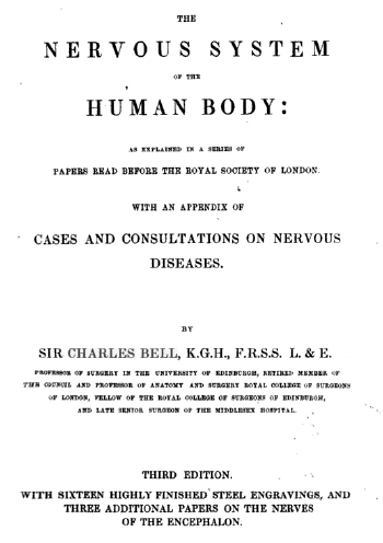
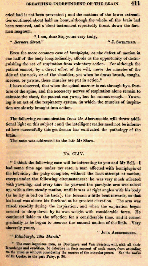
In 1844, the following case was reported in "The
nervous system of the human body as explained in
a series of papers read before the Royal Society
of London" by the Scottish physician John
Abercrombie
(1780-1844), an author well known for his exact
observations and reports of their pathology : "I
had some time ago under my care, a man affected
with hemiplegia of the left side, the palsy
complete, without the least attempt at motion,
except under the following circumstances: he was
very much affected with yawning, and every time
he yawned the paralytic arm raised up, with a
firm steady motion, until it was at right angles
with his body (as he lay in bed on his back),
the forearm a little bent inwards, so that his
hand was above his forhead at its greatest
elevation. The arm was raised steadily during
inspiration, and when the expiration began
seemed to drop down by its own weight with
considerable force. He continued liable to the
affection for a considerable time, and it ceased
gradually as he began to recover the natural
motion of the limb." (Bell, 1844)
Using recent case reports collected from two
differents sites in France and Switzerland, we
aim to present this curious phenomenon, which
involves the combination of involuntary motion
in a paralyzed upper limb with yawning. We
propose afterward a pathophysiological
hypothesis.
Case
reports.
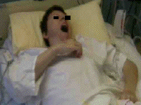
Case 1 (Montreux)
A 49-year-old lady with no history of
vascular disease or recognized risk factors,
except a long history of migraine with aura and
cigarette smoking, developed a fluctuating
hemiplegia on the left side, shortly after
waking up. Weakness predominated in the upper
limb, with moderate dysarthria. Sensory testing
was unremarkable, and no associated neurologic
dysfunction was found on examination.
Diffusion-weighted MR showed a small infarct in
the posterior limb of the right internal
capsule, with associated marked leukoaraiosis.
Blood pressure was normal, as were cardiac
investigations and extra/intracranial MR
angiography. A positive mutation for CADASIL was
found (her father had had strokes and dementia).
After 2 days, the patient reported that while
she was unable to move the left upper limb on
purpose, it raised every time that she yawned,
up to the level of her breast, when she was in a
sitting position. This lasted for a few seconds
and was not associated with specific movements
of the hand and fingers. The phenomenon could be
demonstrated in front of external observers, and
disappeared while motor recovery developed over
the following two weeks.
Case 2 (Montreux)
A 73-year-old gentleman, treated for high
blood pressure and cholesterol, developed
sensorimotor hemiparesis on the right side,
without cognitive changes or aphasia. Initially,
he was completely paralyzed in the upper limb,
but during episodes of yawning, the same limb
would move upwards for about 30 cm, remain still
for a couple of seconds, and then slowly return
to its primary position. This was mainly noted
when the patient was sitting in a wheelchair,
and disappeared after one week, while the
patient recovered some voluntary motricity in
the upper limb, with ataxia. MRI showed a small
infarct in the anterior limb of the left
internal capsule, suggesting lacunar infarction.
An occlusion of the V4 segment of the left
vertebral artery was present, but no large
artery disease was found in the anterior
circulation, and cardiac tests showed no embolic
source.
Case 3 (Poitiers)
A right-handed, 71-year old retired postman
and non-smoker was being treated for
non-insulin-dependent diabetes and
hypercholesterolemia, without hypertension. In
November 1996, he presented with an isolated
motor deficit in the left leg, resolving in 24
hours. The assessment of this transient ischemic
attack revealed bilateral 40%-50% stenosis of
the internal carotid arteries, with
non-ulcerated atheromatous plaques at the right
carotid bifurcation with inhomogeneous echo
structure, without thrombi. A few days later,
magnetic resonance imaging (MRI) of the brain
showed no residual lesions. The echocardiogram
was normal. The patient was prescribed 250 mg of
aspirin per day. In September 1999, sudden onset
of rotatory vertigo was rapidly followed by
severe left hemiplegia without sensory
impairment and without nystagmus or hemianopsia.
The brain CT scan was normal upon the patient's
admission. Doppler examination of the neck
vessels produced the same results as previously.
The Holter ECG recording suggested neither
arrhythmia nor ischemia. The transoesophageal
echocardiogram revealed a left intra-atrial
thrombus. The follow-up CT scan secondarily
associated the ischemic-appearing hypodensity in
the right semioval center and right lenticular
nucleus with leukoaraiosis. The diagnosis was
embolic occlusion of the lenticulostriate
branches of the right middle cerebral artery.
The patient was put on heparin, with a vitamin K
antagonist as relay treatment. One year later,
the patient still had severe left hemiplegia,
predominantly brachiofacial; spasticity was
significant. During a follow-up visit, he
reported the involuntary lifting of his totally
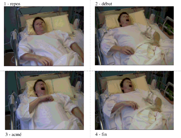
paralyzed left arm during yawning. In 2 to 3
seconds, the arm was raised around 10
centimeters, with adduction and elbow flexion,
at the same time as the patient opened his
mouth. It fell inert once he closed his mouth.
This could occur while he was sitting, standing
or lying down. He had observed this phenomenon
from the onset of his hemiplegia. In June 2002,
three years after the vascular accident, this
involuntary movement of the paralyzed arm during
yawning persisted.
Case 4 (Poitiers)
A right-handed, 53-year-old male bank
employee was known to be a carrier of activated
protein C resistance (Factor V Leiden, treated
with a vitamin K antagonist). This hemostatic
disorder had already caused phlebitis in his
right leg in 1999 and 2000. In addition to this
thrombosis risk factor, he had a high blood
cholesterol level of 2.4 g/L and had smoked
around 15 cigarettes a day since age 14. Between
May 27 and June 2, 2001, he had a series of
alternating transient ischemic attacks,
characterized by predominantly brachiofacial
motor deficits and requiring hospitalization.
The carotid Doppler examination and the
echocardiogram were normal; the patient's blood
pressure was also consistently normal. On June
6, 2001, heparin was stopped for 4 hours to
obtain an angiogram of the supra-aortic
arteries. During this period, a severe left
hemiplegia with sensory deficit and without
hemianopsia developed rapidly and did not
resolve. The brain CT scan revealed an infarct
in the right caudate nucleus and right semioval
center. The angiogram revealed dolichoectasia of
the C2 and C3 portion of the right carotid
siphon, and a thrombus in the middle cerebral
artery at the origin of the lenticulostriate
arteries. In the hours following the cerebral
infarction, the patient was very drowsy and
yawned deeply in an abnormally frequent and
repeated way. At the beginning of each yawn, the
left arm lifted involuntarily with flexion of
the elbow and adduction. This gesture lasted for
the duration of the yawn. The muscular force was
so great that the patient's wife, seated next to
him, was unable to counter it.
Case 5 (Poitiers)
A right-handed, 75-year-old male retired
railway worker with alcoholic cirrhosis
presented on November 7, 2001, with
brachiocephalic severe right hemiparesis
resolving in an hour. His blood pressure was
14/8. However, in the days following, his
systolic blood pressure was highly variable with
several peaks above 20. Because he was living in
a geriatric facility, he did not undergo a
Doppler examination. The brain CT scan was
normal. He received one injection of Enoxaparin
40mg per day. On November 10, 2001, a
brachiocephalic right hemiplegia developed,
associated with lingual paresis, Broca's aphasia
and alliesthesia in the right arm. The patient's
blood pressure was 22/10. The Doppler
examination found left and right internal
carotid lesions which were identical in
appearance and consisted of irregular calcified
atheromatous plaques. The lesions measured 1.5
cm in height and were non-ulcerated without
adherent thrombi, with 50% stenosis, without
hemodynamic repercussions. The vertebral
arteries were normal. The echocardiogram showed
a dilated left atrium, concentric left
ventricular hypertrophy, moderate mitral
insufficiency and an absence of intra-atrial
thrombi. The ECG showed normal sinus rhythm. The
CT scan performed on November 19 (D+9) revealed
an infarct in the left internal capsule and left
lenticular nucleus, without signs of hemorrhage,
without mass effect. The ischemia resulted from
a thrombus in the middle cerebral artery at the
level of the lenticulostriate arteries. From the
onset of the patient's accident, an increased
frequency of yawning was noted. With each yawn,
the right arm lifted with adduction and elbow
flexion, falling as soon as the patient closed
his mouth.
A right-handed, 35-year-old housewife and
smoker presented during the 7th month of her
third pregnancy with superficial calf vein
phlebitis (June 2003). She received a daily
injection of Enoxaparin 40mg for one month. On
October 20, 2003, two months after her delivery,
under low-dose combined oral contraception, she
rapidly developed an inhabitual, diffuse,
intense headache resistant to all the peripheral
analgesics tried. She did not consult a
physician. On October 22, 2003, her husband
found her lying on the ground, paralyzed and
aphasic. Upon her admission to the emergency
room, massive complete right hemiplegia was
noted along with total aphasia, bilateral
mydriasis with pupils showing little
responsiveness, agnosia, an absence of nuchal
rigidity, cerebellar syndrome and sensory
impairment. The patient was very drowsy and
showed a clear automatic-voluntary dissociation.
Blood pressure was 12/7 and cardiac rhythm was
sinusal. The CT scan performed on admission was
normal without subarachnoid hemorrhaging. The
Doppler examination was normal on the right. On
the left, there was complete occlusion of the
internal carotid, with no distal flow. The
transoesophageal echocardiogram was normal. A CT
scan performed in November 2003 (D+20) and
transmitted via teleradiology showed a complete
middle cerebral artery territory infarct and
contrast uptake in the left
frontotemporoparietal cortex. The angiogram
showed fibromuscular dysplasia and left carotid
dissection, responsible for the complete MCA
territory infarct with cortical and subcortical
involvement. From the initial phase of complete
flaccid hemiplegia, every time the patient
yawned, her completely paralyzed right arm
lifted with elbow flexion and moved adductively
towards her face, before falling heavily when
she closed her mouth.
Discussion.
Altough being probably unaware of the
publication of J. Abercrombie in 1844, D
Liégey of Rambervilliers in France
reports in the same manner, in1851, the
following case published in the Gazette
médicale de Strasbourg: "A man recovering
from repetitive attacks of hemiplegia was
overcome by very disagreeable yawning which
occurred every day at the same time. No less
remarkable, whenever this yawning occurred, it
was accompanied by a convulsive movement which
raised the patient's previously paralyzed arm.
The movement produced a painful sensation in the
limb and could only be stopped by the patient
strongly grasping the arm with the opposite
hand."
Hence, since the early 19th century, a few
cases of the movement of a paralysed arm during
yawning have been reported : E.
Darwin 1801, J.
Albercrombie & A. Gendrin 1835,
Liegey 1851 &
1879, Ogle
1863, R.
Trautman 1901, H.
Thomson 1903, M.
Bertolotti 1905, FM.
Walshe 1923, J
Purves-Stewart 1931, A.
Heusner 1946, G.
Mulley 1982, A.
Lanari 1983, H.
Wimalaratna 1988, O.
Blin 1994, E
Louwerse 1998, R.
Töpper 2003. The etiology is either an
ischemic or hemorrhagic vascular accident or a
bulbar form of amyotrophic lateral sclerosis; in
an earlier case, brainstem tuberculoma was the
cause. Based on these cases and the literature
review, this hemiplegia-associated movement
shows no lateral preference and may appear from
the accident's onset during the flaccid phase,
or later, during the spastic phase. The reports
mention either abduction or adduction of the
arm. While the paralysis leaves the arm
immobilized in semi-extension alongside the
patient's body, we have observed the arm to move
outwards away from the thorax (abduction),
followed by elbow flexion bringing the hand
towards the sternum (adduction). This movement
is strictly concomitant with the yawn; the arm
falls inert once the yawn ends. No simultaneous
movement is observed in the leg. The arm's
movement is totally involuntary and
uncontrollable. It coincides with the paralysis
and tends to disappear if there is motor
recovery.
From 1988 on, the case reports have
localized the lesion responsible for the
paralysis. Two locations appear closely linked
to this clinical picture. The first is a lesion
in the middle cerebral artery territory,
particularly the lenticulostriate branches,
leading to infarction in the internal capsule
and lenticular nucleus (paleostriatum) or
caudate nucleus (neostriatum) (Bladin and
Berkovic, 1984). The second is a pontomedullary
lesion, as caused by tuberculoma in a
14-year-old girl with Millard-Gübler
syndrome (Bertolotti, 1905), and also reported
in a series of bulbar amyotrophic lateral
sclerosis cases (Louwerse, 1998) and in a case
of basilar artery thrombosis resulting in
right-sided pontine infarction (Töpper,
2003).
Hemiplegia and associated
movements.
At the end of the 19th century, neurologists
were already discussing the movements associated
with hemiplegia. Vulpian (1866) employed the
term synkinesis for "movements which take place
involuntarily in one part of the body as
voluntary or reflex movements are carried out
elsewhere." In pyramidal syndromes in general
and hemiplegias in particular, synkinesis
produces involuntary movements in paralyzed
muscles concomitant with voluntary movements in
healthy muscles. Classically, there are several
types: global synkinesis, which is merely an
exaggeration of the contraction on the paralyzed
side during an effort on the healthy side;
coordination synkinesis, when the voluntary
contraction of the healthy muscle results in the
involuntary contraction of the paralyzed
synergistic muscles; and imitation synkinesis,
when a voluntary movement on the healthy side
triggers an involuntary attempt at the same
movement on the paralyzed side.
De Buck (1899) also described movements
associated with hemiplegia: "Surprisingly
enough, they extend even to the extremities of
organs not under voluntary control. These
processes accompany not only voluntary
movements, but also reflex movements &endash;
yawning, sneezing, etc." A. Souques wrote about
hemiplegia in Pratique
Médico-Chirurgicale (Brissaud, Pinard and
Reclus, 1907) re-examined this distinction: in
addition to synkinesis which only occurs in the
spastic phase, "associated reflex movements
coincide with a cough, a yawn, primarily
affecting the upper limb" and may be observed
during the initial flaccid phase. Working in
Turin, Bertolotti (1905) made the same
distinction. And Brissaud wrote in his lecture
dated December 1, 1893: "Spasmodic yawning is
also seen with a certain frequency in
hemiplegics. Not merely a superficial symptom
amongst the spasmodic elements of epilepsy
(Féré) or hysteria (Charcot,
Gilles de la Tourette, Guinon and Huet), it
indeed seems to arise directly from organic
lesions...." We agree with the disciples of
J.-M. Charcot on this point, and do not support
using the term synkinesis to describe this
involuntary raising of the arm during
yawning.
In vertebrates, ethologists describe
pandiculation or Rekel-Syndrom as the
association of yawning with a stretching of the
trunk and four limbs (Selbach and Selbach,
1953). Bertolotti (1905) and Blin (1994), each
reporting similar cases to those described
above, use the term pandiculation or
hemipandiculation. Neither the description of
the arm's involuntary, non-stretching movement,
nor the absence of stretching in the leg on the
same side, support the use of this term.
De Buck (1899) wrote: "Parakinesia appears
to differ from synkinesia by the fact that the
relationship between the idea and the act in the
latter is perfectly preserved, involving only a
simple diffusion of the nervous influx at the
motor centers, owing to the need for a much
stronger signal to produce a useful effect; in
parakinesia, the relationship between the idea
and the movement appears disrupted." We have
therefore proposed the term parakinesia
brachialis oscitans. Parakinesia is an abnormal
movement that acts as a parasite, caricature or
replacement of a normal movement, in this
instance of the arm (brachial). Oscitans comes
from the Latin oscitantis, meaning "which yawns"
and mentioned in early writings on fever
(Thomson, 1827).
Yawning.
Yawning is phylogenetically ancient,
resembling to what ethology appoints a
maintenance behavior. Highly stereotypical, it
is observed in cold-blooded and warm-blooded
vertebrates, from reptiles with rudimentary,
"archaic" brains to human primates, in water,
air and land environments. The ethology,
neurophysiology and neuropsychology literature
associates yawning with wake/sleep rhythm
fluctuations, eating, and sexuality, where it
externalizes a group of vigilance-stimulating
mechanisms and attests to the hypothalamus'
central role in homeostasis (Aubin and Garma,
1988; Daquin et al., 2001; Walusinski and
Deputte, 2004). Yawning is recognizable in
ultrasound images from the
14th week of pregnancy, and like the
appearance of oromandibular movements and
swallowing, it signals functional maturation of
the brainstem and basal ganglia, whereas the
extension of the frontoparietotemporal cortex
continues through the 24th week (Abadie et al.,
1999). No brain structure has ever been
identified as the yawning center. Clinical and
pharmacological evidence indicates that the
hypothalamus &endash; particularly the
paraventricular nucleus &endash; plays a key
role in yawning, as well as medullary and
pontine regions, with connections towards the
frontal region in human primates and towards the
cervical spine. The muscles that contract during
yawning are supplied by cranial nerves V, VII,
IX, X, XI and XII, cervical nerves C1-C4
(phrenic nerve) and the dorsal nerves
innervating the intercostals, which play an
accessory role in breathing (Chouard and
Bigot-Massoni, 1990).
Provine et al. (1987) showed that yawning
has no effect on arterial oxygenation. Hence,
the old paradigm, which from the time of
Hippocrates to the mid-20th century posited this
behavior as a reflex to increase brain oxygen
supply, must now be replaced with a
neuromuscular paradigm involving subcortical
control. The hypothalamic paraventricular
nucleus contains the cell bodies of oxytocin
neurons projecting to the hippocampus and to the
reticular formation and locus ceruleus in the
brainstem. When these neurons are stimulated by
dopamine, excitatory amino acids or oxytocin
itself, they trigger yawning by releasing
oxytocin in these various subcortical
structures. This oxytocinergic activation is
inhibited by opioids, which prevent yawning. The
activation or inhibition of these oxytocin
neurons is correlated with the activity of
paraventricular nitric oxide synthase. Other
neuronal systems are involved as modulators.
Serotonin, estrogens, testosterone and
hypocretin play a role either in the
paraventricular nucleus or the motor nuclei of
the brainstem and spinal cord, with the final
executive pathway controlled by acetylcholine
(Argiolas and Melis, 1998; Blin, 1996;
Sato-Suzuki et al., 2002).
Attempt at a
pathophysiological explanation.
The release of subcortical structures from
cortical inhibition is classically proposed to
explain certain automatic or reflex activities
occurring experimentally in decorticate animals
or following stroke in humans. This is the case
for palatal myoclonus, or palatal tremor
(Lapresle, 1984), caused by destruction of the
central tegmental tract, allowing uncontrolled
activity in the olivary body and, in turn, the
palate's rhythmic movement. This amounts to a
reemergence of the structure's phylogenetically
branchial function.
Another example is the syndrome of Foix,
Chavany and Marie (1926), or biopercular
syndrome, where the automatic activities of
emotional expression, swallowing and yawning
remain intact even though there is total
faciopharyngoglossomasticatory paralysis. The
automatic activity depends on the brainstem and
is preserved, whereas the voluntary control of
the prerolandic cortex and the supplementary
motor area is interrupted by a biopercular
infarct. One MRI case study showed this
automatic-voluntary dissociation as a
corticobulbar disconnection (Ghika and
Bogousslavsky, 2003).
A final example is locked-in syndrome, in
which there is total paralysis and patients may
only retain voluntary control over eye movement.
However, involuntary facial expressions of pain
are preserved, along with yawning and loud
crying. The corticospinal or pyramidal tract is
completely interrupted at the pons. The
preserved activity of extrapyramidal and
cerebellar structures and of archaic spinal
automatisms explains the automatic-voluntary
dissociation (Bauer et al., 1980).
Could such a mechanism of
cortical-subcortical dissociation explain the
synchronization between the raising of the arm
and the respiratory cycle of the yawn, due to
diaphragmatic extension?
The study of the relationship between
respiratory cycles and movement was initiated by
Marey (1892) through his invention of
chronophotography, a precursor to the cinema. He
had already shown that the breathing cycle of a
running dog, a galloping horse and a flying gull
was based on stride or wingbeat. This
synchronicity allows optimal adaptation between
oxygenation and muscular work, but the resulting
piston effect of the diaphragm's movement also
plays an important role in bodily and
aerodynamic equilibrium. Energy transfers
between trunk muscles and those used for
breathing improve the energetic yield of running
(Bramble and Carrier, 1983; Hsueh-Tze, 1997;
Viala, 1986). Humans maintain partial coupling
with arm-swinging during walking, a sign of
extrapyramidal activity (Persegol et al., 1991;
Bernasconi and Kohl, 1993).
Respiratory automatisms are the result of
several medullary and pontine nuclei working in
concert. Experimental data suggest that
respiratory rhythm generation depends on
pacemaker neurons. These neurons are found in a
small region known as the pre-Bötzinger
complex, ventral to the nucleus ambiguus and to
the emergence of the roots of cranial nerve XII.
The pacemaker-generated rhythm is modulated by
networks of respiratory interneurons,
responsible for its final spatiotemporal
organization. Inspiratory and expiratory neurons
have been described according to the respiratory
phase concurrent with their firing time. The
rhythmic signal from these neuronal interactions
is then distributed, according to a precise
spatiotemporal pattern, to medullary and spinal
motor neurons, thereby ensuring the motor
innervation of the upper airway and respiratory
muscles (Bianchi et al., 1995; Duffin and Ezure,
1995).
Adjacent to these nuclei, the lateral
reticular nucleus projects to the cerebellum and
plays a role in the sensory-motor coordination
of the limbs. This corresponds to the
spinoreticulocerebellar pathway. Experiments in
the cat have shown that when the nuclei in the
respiratory rhythm complex are firing, the
lateral reticular nucleus itself has a
rhythmical and synchronous activity,
particularly when limb extensor muscles are
contracted during stretching (Arshavsky, 1978
and 1986; Ezure and Tanaka, 1997; Richard and
Waldrop, 1989). The dual role of the
intercostals and the diaphragm in posture and
locomotion on the one hand, and in breathing on
the other (not to mention phonation), requires
not only somatomotor coordination, but also
neurovegetative coordination, especially of
cardiovascular functions. In the cat, Schomburg
et al. (2003) showed sympathetic modulation of
cardiolocomotor and respiratory activity with a
spinopontine feedback loop extending to the
hypothalamus.
Cardiorespiratory adaptation to effort
involves the autonomic nervous system,
particularly the hypothalamus and the pituitary
gland, with regulation of blood pH, PaCO2 but
also satiety and vigilance (Waldrop et al.,
1986; Yeh et al., 1997). These homeostatic
mechanisms, dependent on anatomically similar
subcortical structures always in close relation
and regulated by identical neurotransmitters,
are linked to those controlling the rhythms of
wake/sleep, satiety and sexuality, i.e. those
involved in triggering yawning (Salin-Pascual et
al., 2001; Walusinski and Deputte, 2004).
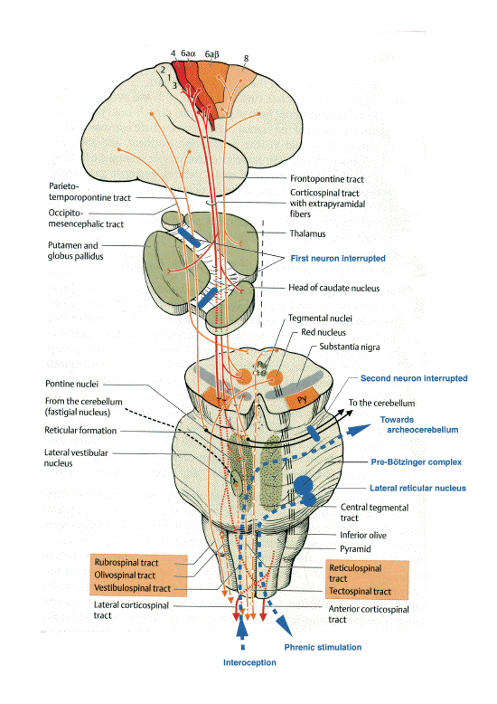
The extrapyramidal motor system has a key
influence on the spinal, brainstem and
cerebellar motor circuits as well as the motor
cortex itself. Its neurons connect the cerebral
cortex to the cerebellum, forming the
corticopontocerebellar tract of the
neocerebellum, whose phylogenetic development
mirrored that of bipedality (Llinas and Sotelo,
1992). The cerebellum receives a copy of all
motor impulses from the cerebral cortex through
this tract (Schweighofer et al., 1998). This
corticopontine pathway passes through the
internal capsule on each side of the pyramidal
tract. The lesion responsible for parakinesia
brachialis oscitans is most often found at this
level, involving damage to the first neuron and
interrupting the corticospinal and
corticonuclear pathways, but also the
extrapyramidal corticostriate, corticorubral,
corticonigral and corticoreticular pathways (see
figure above).
At the level of the pons, the fibers form
synapses in the pontine nuclei whose axons
constitute the second neuron projecting to the
cerebellum (middle cerebellar peduncle). The
cases reported by M. Bertolotti (1905), L.
Louwerse (1998) and R. Töpper (2003) show
that a lesion at this level may also cause
parakinesia brachialis oscitans (fig. 1).
The paleocerebellum receives signals from
the spine via the dorsal and ventral
spinocerebellar tracts. The two pathways
transmit proprioceptive impulses from the
peripheral system, i.e. from muscle spindles and
Golgi tendon organs in the muscles of the limbs,
trunk and diaphragm. Efferents from the
paleocerebellum, after being relayed by the
emboliform nuclei and the fastigial nucleus,
project to the red nucleus via the superior
cerebellar peduncle. In this way, the
paleocerebellum controls antigravity muscles,
determines the appropriate muscle tone for
walking, participates in the synergistic
coordination of agonist and antagonist muscles
necessary for this task, all while coordinating
the motor control of the diaphragm. It also
sends out ascending signals which project to the
centromedian nucleus of the thalamus, then to
the caudate nucleus and the putamen, thereby
providing a connection with the extrapyramidal
system (Duss, 1998).
Like normal walking, pandiculation &endash;
the generalized stretching of the trunk, limbs
and diaphragm &endash; requires the
uninterrupted functional activity of the
corticoneocerebellospinal and the
spinoarcheocerebellothalamic pathways, coupling
the pyramidal and extrapyramidal control of the
entire musculature.
Interruption of the corticospinal tract,
whatever the etiology, causes paralysis in the
arm, preventing voluntary motion. In certain
cases, the cortioneocerebellospinal pathway is
also interrupted. However, the proprioceptive
loop conducting signals between the motor
anterior spinal horn, the paleocerebellum and
the lateral reticular nucleus (ventral
spinocerebellar tract) remains functional.
During yawning, the strong contraction of
respiratory muscles represents a proprioceptive
signal. An antidromic stimulation from the
respiratory nuclei to the anterior spinal horn
has been experimentally demonstrated in the cat
(Ezure and Tanaka, 1997). It might be
speculated, albeit boldly, that parakinesia
brachialis oscitans is a movement of the arm
(akin to the swinging coupled with respiration
during walking) secondary to an incoming motor
signal in the anterior spinal horn from C4 to
C8, originating in the lateral reticular nucleus
and traveling through the extrapyramidal
pathways of the archeocerebellum (Waldrop et
al., 1986).
This mechanism is consistent with the
absence of movement in the leg (corticospinal
interruption) and with the possibility of
occurrence in the flaccid as well as the spastic
phase. A preserved corticoneocerebellar pathway
would prevent such a movement. Therefore, the
requisite condition for parakinesia brachialis
oscitans would be corticonuclear, corticospinal
and corticoneocerebellar interruption, whereas
the spinoarcheocerebellar pathway would remain
functional. Assuming that yawning is the
exteriorization of a homeostatic mechanism
regulating the vigilance systems in the
hypothalamus, repetitive yawning, which is
frequently observed during stroke, would appear
to stimulate arousal mechanisms. When a lesion
in the extrapyramidal system prevents the
modulating activity of the corticoneocerebellar
pathway, structures that are phylogenetically
more primitive may allow recovering movement in
the arm during the diaphragm's maximal
ampliation, as occurs during yawning.
The central nervous system in vertebrates
follows a common organizational pattern and
shows gradually increasing complexity with
higher and higher levels of independence and
functionality. The American neuropsychiatrist P.
MacLean (1985) proposed a model of the nervous
system's functional organization based on the
study of its phylogenesis. At the base of this
model is the ancestral "reptilian" brain
(brainstem and basal ganglia), where yawning
originates. The next level is the
"paleomammalian" brain (limbic system) shared by
all mammals. This is the synaptic and hormonal
interface, where emotive yawning in monkeys is
localized. Finally, a "neomammalian" brain
comprises the top layer, characterized by
cortical development in humans, particularly of
the frontal lobes, where "contagious" yawning
occurs (Walusinski and Deputte, 2004). If these
functional levels become disconnected, as
happens in certain stroke localizations,
functions may reappear that are normally
inhibited by a phylogenetically more recent and
functionally more sophisticated structure. In
this way, human pathology reveals that the
coordination and regulation of body temperature,
breathing, locomotion and vigilance has been
perfected over time, with ever-increasing
complexity and precision, from reptiles to
primates.
Argiolas
A, Melis M.R. The neuropharmacology of
yawning Eur Pharmacol; 1998; 343; 1; 1-16
Arshavsky
YI. et al Messages convey by
spino-cerebellar pathways during scratching in
the cat Brain Res 1978; 151; 479-506
Arshavsky YI. et al Cerbellum and rhythmical
movements Springer Berlin 1986
Aubin H.J,
Garma L Le bâillement Psychiatrie et
Psychobiologie; 1988; 3; 275-286
Bauer G. et al
Involuntary motor phenomena in the locked in
syndrome J Neurology 1980; 223; 191-198
Bernasconi
P, Kohl J. Analysis of coordination between
breathing and exercise rhythms in man J
Physiology 1993; 471; 693-706
Bertolotti
M. Etude sur la pandiculation automatique
des hémiplégiques Rev Neurol 1905;
2; 19; 953-959
Bianchi
AL, Denavit-Saubié M, Champagnat J.
Central control of breathing in mammals :
neuronal circuitry, membrane properties, and
neurotransmitters. Physiological Reviews 1995;
75; 1-45
Blin
O. Le bâillement en
neuropsychopharmacologie clinique Lettre du
pharmacologue 1996; 10; 217-219
de
Buck Classification des mouvements anormaux
associés à
l'hémiplégie Rev Neurol 1899; 6;
361-374
Duan
Y et al Cardiorespiratory components of
defense reaction elicited from paraventricular
nucleus Physiol Behav 1997; 61; 2; 325-330
Ezure K,
Tanaka I. Convergence of central respiratory
and locomotor rhythms onto sigle neurons of the
lateral reticular nucleus Exp Brain Res 1997;
113; 230-242
Foix C, Chavany
JA, Marie J. Diplégie
facio-linguo-masticatrice d'origine
cortico-souscorticale sans paralysie des membres
Rev Neurol 1926, 33, 214-219
Fugl-Meyer
AR,, Linderholm H et al. Restrictive
ventilatory dysfunction in stroke: its relation
to locomotor function Scand J Rehabil Med 1983;
Suppl 9; 118-124
Furtado D.
Provocation spinale d'un réflexe de
bâillement. Rev d'oto neuro
ophtalmologie.1951;23(1):
Ghika
J, Bogousslavsky J. Dissociated preservation
of automatic-voluntary jaw movements in a
patient with biopercular and unilateral pontine
infarcts Eur Neurol 2003, 50, 185-188
Heusner A P
Yawning and associated phenomena Physiological
Review 1946; 25; 156-168
Hsueh-tze
L. Mechanical links between locomotion and
beathing: can you breath with your legs ? News
Physiol. Sci. 1997; 12; 273-278 télécharger
PDF
Lanari A,
Delbono O The yawning and stretching sign in
hemiplegics Medicina (B Aires) 1983; 43; 3;
355-356
Lapresles
J La voie dento-olivaire: sa mise en
évidence, son trajet, sa signification
Bull Acd Nat Med 1984, 168, 3-, 336-341
Liecey Nouvelle
observation de bâillement convulsif
périodique Le Courrier Médical
1879; 29; 334-336
Liégey
Deux observations de bâillements
intermittents Gazette médicale de
Strasbourg 1851; p118-119
Louwerse
E Forced yawning as a pseudobulbar sign in
amyotrophic lateral sclerosis J Neuroscience
Research 1998, sup, 392
MacLean P
Evolutionay psychiatry and the triune brain;
Psychological Medecine 1985; 15; 219-221
Marey
EJ La photographie du mouvement G.
Carré ed Paris 1892, 97p (le
musée)
Mignot E., et al Sleeping with the
hypothalamus Nature Neuroscience 2002; 5;
1071-1075
Morin
D, Viala D Coordinations of locomotor and
respiratory rhythms in vitro are critically
dependent on hindlimb sensory inputs. J Neurosci
2002; 22; 11; 4756-65. télécharger
PDF
Mulley G
Assoctiated reactions in the hemiplegic arm
Scand J Rehab Med 1982; 14; 117-120
Ogle
JW Arm rising during yawning The Medical
Times and Gazette. 28 fébruary 1863 p
213
Persegol
L, Jordan M et al. Evidence for central
entrainment of the medullary respiratory pattern
by the locomotor pattern in the rabbit. Exp
Brain Res 1988; 71; 1; 153-62
Persegol
L, Jordan M et al. Evidence for the
entrainment of breathing by locomotor pattern in
human. J Physiol (Paris) 1991; 85; 1; 38-43
Pierre
Marie La Pratique Neurologique
Hémiplégie, mouvements
associés Masson 1911, p477-480
Pierre
Marie et André Léri Mouvements
involontaires dans les membres paralysés
1911 Nouveau Traité de médecine et
de thérapeutique Brouardel, Gilbert,
Thoinot JB Baillière Ed 1911
p283-291
Quoirin
E. Elévation involontaire du membre
supérieur chez
l'hémiplégique lors d'un
bâillement Thèse doctorat en
médecine Poitiers 2002
Richard
CA, Waldrop TG et al. The nucleus
reticularis gigantocellularis modulates the
cardiopulmonary responses to central and
peripheral drives related to exercise Brain Res
1989; 482; 1; 49-56
Romberg
MH A manual of the nervous diseases of man.
Berlin - London 1853
Rosenbaum D. Human Motor Control 300 p
Academic Press 1990
Salin-Pascual
R. et al Hypothalamic regulation of sleep
Neuropsychopharmacology 2001; 25; S21-S27
Sato-Suzuki
I, I Kita, Y, Seki, M Oguri, H Arita.
Cortical arousal induced by microinjection of
orexins into the paraventricular nucleus of the
rat Behavioural Brain Research 2002; 128;
169-177
Schiller
F Yawning ? J History of the Neurosciences
2002; 11; 4; 392-401
Schomburg
ED et al Rhytmic phrenic, intercostal and
sympathetic activity in relation to limb and
trunk motor activity in spinal cats Neuroscience
Research 2003: 46; 229-240
Selbach C, H
Selbach Das Räkel-Syndrom als
Wirkungsfolge eines biologischen Regelsystems.
Monatschr.f. Psychiat u Neurol; 1953; 125;
671-82.
Thomson HC
Associated movements in hemiplegia : their
origin and physiological significance Brain
1903; 26; 515-523
Töpper
R, Mull M, Nacimento W Involuntary
stretching during yawning in patients with
pyramidal tract lesions: further evidence for
the existence of an independent emotional motor
system European J Neurology 2003; 10;
495-499
Trautmann R
Le bâillement Thèse Bordeaux;
1901-02; N° 40; 86 pages
Viala
D Evidence for direct reciprocal
interactions between the central rhythm
generators for spinal "respiratory" and
locomotor activities in the rabbit Exp Brain Res
1986; 63; 2; 225-32
Vulpian
A Leçons sur la physiologie
générale et comparée du
système nerveux faites au Museum
d'histoire naturelle. Rédigées par
E.Brémond. Paris, Baillière,
1866
Vulpian
A Maladies du système nerveux Paris,
Doin, 1879
Waldrop
TG et al Control of respiration by the
hypothalamus and feedback from contracting
muscles in cats Respir. Physiol 1986; 64;
317-328
Walusinski O,
Deputte BL Le bâillement :
phylogenèse, éthologie,
nosogénie Revue Neurologique
2004;160(11):1011-1021
Walshe FMR
On certain tonic or postural reflexes in
hemiplegia with special reference to the so
called "associated movements". Brain,
1923;46:1-37
Wimalaratna
HS, Capildeo R. Is yawning a brainstem
phenomenon ? a stroke patient who stretched his
hemiplegic arm during yawning Lancet 1988; 1;
8580; 300
Yeh
E et al The paraventricular nucleus of the
hypothalamus influences respiratory timing and
activity in the rat Neurosci Let 1997; 232;
63-66
The nervous system of the human body as
explained in a series of papers read before the
Royal Society of London with an appendix of
cases and consultations on nervous
diseases.
Charles Bell - London. Henry Renshaw Ed.
1844. p411
Schematic representation of the
extrapyramidal tract and the localizations of
lesions responsible for parakinesia brachialis
oscitans. (adapted from Duus' Topical Diagnosis
in Neurology, Thieme ed. 2005)