Involuntary
stretching during yawning in patients with
pyramidal tract lesions: further evidence for
the existence of an independent emotional motor
system
R. Topper, M. Mull, W. Nacimiento
Department of Neurology and
Neuroradiology, Aachen Technical
University,Germany
Introduction : Yawning is a
stereotyped behavioural pattern which begins
with an inspiration associated with marked
dilatation of the pharynx. At the peak of
inspiration there are associated facial
movements and the final part of yawning is
passive expiration. Yawning is accompanied by
lacrimation, salivation, and reflex
vasoconstriction in the skin (Barbizet,
1958; Appenzeller, 1969). Ultrasound has
revealed that yawning occurs even in fetuses in
utero (Egerman and Emerson, 1996).
Not much is known about the function of
yawning. Folk wisdom commonly associates yawning
with drowsiness and boredom. The few scientific
studies which have attempted to unravel the
physiological function of yawning have not
succeeded in formulating a convincing
hypothesis. The popular opinion, that yawning is
a respiratory manoeuvre to increase oxygenation
and decrease C02 in the blood has not been
verified in normal subjects. In an experimental
setting the subject had either to breathe 100%
02 or a gas mixture with higher than normal
levels of C02. In both conditions the frequency
of yawning remained unchanged, which makes it
unlikely that yawning serves any specific
respiratory function (Provine
et al., 1987). Others have argued that
yawning may serve as a paralinguistic signal of
drowsiness in man. An argument in support of
this hypothesis is the fact that yawning is
contagious. Seeing somebody yawning is a
powerful stimulus to evoke a yawn in the
observer. When analysing group behaviour
ethnologists suggested that yawning may help to
synchronize the physiological and behavioural
state of a group (Eibl-Eibesfeld, 1975).
In humans and animals, yawning is often
accompanied by generalized limb extension. In
laboratory animals a variety of dopaminergic and
cholinergic substances have been found that
induce a stereotyped motor pattern which
consists of yawning, stretching and penile
erection (Dourish
and Cooper, 1990). In humans yawning may
occur without associated limb extension (Provine
et al., 1987). An involuntary stretching of an
otherwise plegic arm is, however, often observed
in neurological patients, for example in
patients with
hemiplegia. Other reflexive associated
movements, also known as synkinesias, have been
observed in plegic limbs. These include raising
of the arm and flexion of the thigh whilst
sneezing or moving the arm during micturition
(Walshe, 1923). The
first detailed study of synkinesias came from
the German neurologist Westphal (quoted in
Zülch and Müller, 1969), who
attributed the reflexive movement of the
otherwise plegic arm to the action of the
ipsilateral, uncrossed pyramidal tract. Whilst
other types of synkinesias, such as mirror
movements, received some attention in the
neurological literature (Zülch and
Müller, 1969), associated movements during
yawning have only rarely been described. There
are a few case reports of stroke patients who
experienced stretching of the arm whilst yawning
(Bauer et al., 1980;
Wimalaratna and
Capildeo, 1988;Blin
et al., 1994), but according to one report
this phenomenon might be rather common: in this
study 31 of 40 patients questioned reported that
their plegic arm moved during yawning (Mulley,
1982). The anatomical pathways which
underlie this involuntary motor response have,
however, yet to be clarified. We report three
patients with radiologically characterized
lesions at différent levels of the
pyramidal tracts who experienced involuntary
movements of the otherwise plegic arm, during
yawning.
Case reports
Patient 1 : A 62-year-old
patient was admitted with a left sided
hemiparesis and a hemianopia to the left. A
brain CT obtained on admission revealed an
infarction in the right posterior artery
territory and early signs of an extensive
infarction in the territory of the right middle
cerebral artery. Consecutive CT scans obtained
over the following days showed an extensive
swelling of the ischaemic brain tissue with a
shift of midline structures to the left. The
ischaemic oedema did not respond to vigorous
medical therapy including hyperventilation and
osmotherapy. An extensive craniotomy was
therefore performed 6 days following the onset
of symptoms. After 12 days of artificial
ventilation the tracheal tube was removed. A CT
scan at that time revealed a complete infarction
of brain tissue in the territory of the right
middle cerebral artery. On neurological
examination the patient had a complete left
sided hemianopia and a left-sided spatial
neglect. There was a complete left-sided
hemiplegia with increased muscle tone,
exaggerated reflexes and a positive Babinski
sign on the left. During ward rounds the patient
reported spontaneous movements of his left arm
during yawning. Due to the severity of his
spatial neglect the associated movements of his
left arin gave hiin a rather strange feeling,
especially when his left arm becarne visible for
him on the right side of his body.
Patient 2 : A 51-year-old man
was admitted to the neurology department because
of a dense hemiparesis of his right arm and leg
with sudden onset during physical exercise. His
medical history was remarkable for uncontrolled
arterial hypertension. On neurological
examination the patient was alert and
orientated. He had a central facial paresis and
a complete right sided hemiplegia. The speech
was dysarthric, but there was no aphasia. A
brain CT scan obtained on the day of admission
showed a haemorrhage centred in the left
thalamus which extended into the posterior
portion of the internal capsule. The diagnosis
of a hypertensive intracerebral haemorrhage was
made. A neurosurgical intervention was discussed
but it was decided to treat the patient
conservatively. Approximately 2 weeks after the
stroke a neurology resident observed involuntary
movements of the patient's plegic arm during
yawning. These movements consisted of a tonic
abduction of the arm at the shoulder and an
extension of the forearm and the fingers. The
appearance of these associated movements was
consecutive with the appearance of spastic
muscle tone.
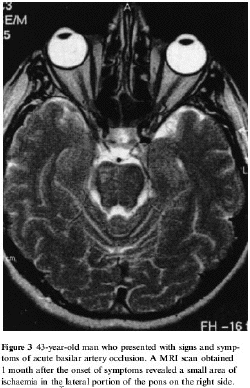
Patient 3 : The last patient
described in this report was a 43-year old man
who was admitted with a basilar artery
thrombosis. He presented with increasing
clouding of consciousness, anisokoric pupils,
restricted extraocular movements of both eyes
and a left sided hemiplegia. An initial brain CT
was normal. An emergency cerebral angiography
revealed a thrombotic occlusion of the basilar
artery in its upper portion. Intra-arterial
application of 90 mg rtPA achieved an almost
complete recanalization of the basilar artery.
After completion of the thrombolytic therapy the
patient remained on artificial ventilation for
the next 24 h. Following extubation the
neurological examination revealed a bilateral
gaze evoked nystagmus, dysarthria, a complete
left sided hemiplegia and sensory disturbances
of the left side of the body. A cranial MRI
obtained 14 days after basilar artery occlusion
showed a circumscribed right sided lesion in the
middle portion of the pons extending to the
cerebellar peduncle
(Fig). As
soon as 4 days after the stroke a nurse on the
neurological intensive care unit noted
spontaneous movements of his plegic left arm
during yawning. This observation could be
confirmed by the patient who noted an abduction
and extension of his left arm every time he
yawned. When he imitated a yawn no associated
movements could be observed.
Discussion
Associated movements of the plegic arm
during yawning were found in three patients with
pyramidal tract lesions at the level of the
motor cortex, the internal capsule and the pons.
The recovery of movements after a pyramidal
tract lesion is characterized by an initial
phase of flaccid paresis which is followed by
the reappearence of tendon reflexes as well by
the appearence of pathological reflexes such as
the Babinski
sign and exaggerated flexor reflexes.
Synkinesias such as stretching of the arm whilst
yawning typically appear at this stage of the
recovery process, before the reappearance of
voluntary limb movements. From these observation
it may be concluded that stretching during
yawning is an automatic motor pattern that is
usually inhibited in the presence of intact
corticobulbar fibres in man. When the
corticobulbar systems have been injured this
automatic motor pattern appears in a stereotyped
fashion.
So far there have been no convincing
hypotheses concerning the anatomical pathways
which are responsible for the involuntary
movements of the arms in the absence of a
functional pyramidal tract. In recent years
yawning has gained increasing recognition from
behavioural neuroscientists studying the effects
of dopaminergic and cholinergic drugs on the
rodent brain. In animals the yawning response to
the dopamimetic apomorphine is considered a
behavioural consequence of the stimulation of
dopaminergic receptors in the basal ganglia, as
experimental basal gangha lesions are known to
abolish apomorphin-induced yawning (Dourish
and Cooper, 1990). The associated movements
of the extremities during yawning in man
following lesions of the corticospinal tract
have therefore been attributed to an activation
of otherwise inhibited basal ganglia projections
onto brain stem motor centres (Blin
et al., 1994). An intact basal ganglia
projection to the brain stem is not, however, a
prerequisite for yawning with associated
stretches in man. Yawning with associated
movements of the extremities has been observed
in locked-in patients following bilateral
midbrain infarction (Karp and Hurtig, 1974;
Bauer et al., 1980).
In an infant born with an arhinencephalic brain
yawning was also prescrit demonstrating that
yawning is possible in the absence of basal
ganglia structures (Gamper,
1926).
There are reports of patients which indicate
that yawning is integrated in the lower brain
stem. A patient with an extensive glioma in the
anterior part of the pons developed a locked-in
syndrome including complete paralysis of all
facial muscles. Reflexive yawning with typical
innervation of facial and laryngeal muscle was
observed which was not, however, accompanied by
reflexive stretching of the arm. (Gschwend,
1977). The anatomical substrate of the
'cerebral yawning centre' is most likely to be
located in the caudal medulla, where neurones
regulate respiratory activities via their
projections to muscles involved in respiration.
In the cat cells in the lateral reticular
nucleus in the lower medulla adjacent to the
ventral respiratory group have been found to
have both central respiratory and locomotor
rhythms (Ezure and Tanaka, 1997). These cells,
if present in man, might be the anatomical basis
for associated stretching during yawning.
It has been suggested that yawning
represents an integrated discharge of the bulbar
reticular formation corresponding to a
particular level of activity of the reticular
formation (Barbizet,
1958). Following this line of arguments
yawning could be considered as the somatomotor
manifestation of a particular emotional state
characterized by disinterest and sleepiness,
just as laughing is the somatomotor
manifestation of mirth. Both have a distinct
psychosocial significance in indicating either
boredom or joy to others. From clinical
observation it can be deduced that the
integrated discharge of the brain stem neurones
responsible for the stereotyped behavioural
pattern, which corresponds to a particular
emotional state, leads to an activation of
bulbar and spinal motor neurones independent of
the pyramidal tract input necessary for a
voluntary activation of the respective muscles.
Patients with lesions of the pyramidal tract at
various levels who have a facial palsy or a
paralysed arm may show an involuntary
innervation of the face during laughing
(Monrad-Krohn, 1924; Hopf et al., 1992;
Tôpper et al., 1995; Waxman, 1996) or an
involuntary stretching during yawning. Holtstege
has recently introduced the term 'emotional
motor system' into clinical neuroscience
(Holtstege, 1991; Holtstege et al., 1996).
This concept implies that descending fibre
tracts within the brain stem, completely
distinct from those forming the somatomotor
system, are involved in the elaboration of
emotional behaviour. Our observations of
movements of an otherwise plegic limb supports
the concept of an independent emotional motor
system. Careful clinical observation combined
with high resolution brain imaging in patients
with circumscribed brain lesions is therefore a
suitable tool to study the somatomotor
components of emotions, a task which is
difficult to perform in laboratory animals.
Is yawning a
brainstem phenomenon ? Wimalaratana HS,
Capildeo R. A stroke patient who stretched his
hemiplegic arm during yawning
Lancet 1988; 1; 8580;
300
de
Buck Classification des mouvements anormaux
associés à
l'hémiplégie Rev Neurol
1899;6:361-365
Furtado D.
Provocation spinale d'un réflexe de
bâillement. Rev d'oto neuro
ophtalmologie.1951;23(1):
Ghika
J., Bogousslavsky J. Dissociated
preservation of automatic-voluntary jaw
movements in a patient with biopercular and
unilateral pontine infarcts Eur Neurol
2003;50:185-188
Heusner A P
Yawning and associated phenomena Physiological
Review 1946;25:156-168
Lanari A.,
Delbono O. The yawning and stretching sign
in hemiplegics Medicina (B Aires)
1983;43(3):355-35
Liecey Nouvelle
observation de bâillement convulsif
périodique Le Courrier Médical
1879;29;334-336
Liégey Deux
observations de bâillements intermittents
Gazette médicale de Strasbourg 1851;
p118-119
Louwerse E
Forced yawning as a pseudobulbar sign in
amyotrophic lateral sclerosis J Neuroscience
Research 1998, sup, 392
Mulley G.
Assoctiated reactions in the hemiplegic arm
Scand J Rehab Med 1982;14:117-120
Ogle
JW. Arm rising during yawning The Medical
Times and Gazette. 28 february 1863 p 213
Pierre
Marie La Pratique Neurologique
Hémiplégie, mouvements
associés Masson 1911, p477-480
Pierre
Marie et Léri A. Mouvements
involontaires dans les membres paralysés
1911 Nouveau Traité de médecine et
de thérapeutique Brouardel, Gilbert,
Thoinot JB Baillière Ed 1911
p283-291
Quoirin E.
Elévation involontaire du membre
supérieur chez
l'hémiplégique lors d'un
bâillement Thèse doctorat en
médecine Poitiers 2002
Thomson HC
Associated movements in hemiplegia : their
origin and physiological significance Brain
1903;26:515-523
Töpper
R, Mull M, Nacimento W Involuntary
stretching during yawning in patients with
pyramidal tract lesions: further evidence for
the existence of an independent emotional motor
system European J Neurology 2003;10:495-499
Trautmann R. Le
bâillement Thèse Bordeaux; 1901-02;
N° 40; 86 pages
Vulpian A
Leçons sur la physiologie
générale et comparée du
système nerveux faites au Museum
d'histoire naturelle. Rédigées par
E.Brémond. Paris, Baillière,
1866
Vulpian
A Maladies du système nerveux Paris,
Doin, 1879
Walshe FMR
On certain tonic or postural reflexes in
hemiplegia with special reference to the so
called "associated movements". Brain,
1923;46:1-37
Wimalaratna HS,
Capildeo R. Is yawning a brainstem
phenomenon ? a stroke patient who stretched his
hemiplegic arm during yawning Lancet
1988;1(8580):300