Cyclic
variation in the amplitude of a brainstem reflex
during sleep and wakefulness
Chase MH, McGinty DJ, Sterman MB.
In recent years, attention has been drawn to
variations in the amplitude of spinal reflexes
which ocçur during the different stages
of sleep and wakefulness. We were interested in
determining if similar variations take place in
reflex activity at a brain-stem level. For this
purpose we chose as a test reflex the masseteric
monosynaptic reflex. It is our intent to
determine the fluctuations in brainstem reflex
transmission during various behavioral states;
the initial ones studied were sleep and
wakefulness.
A second objective of these studies arose as
the result of experiments in acute preparations
in which we observed complete inhibition of this
reflex and others following orbital-cortical or
basal forebrain stimulation. We are presently
investigating the behavioral conditions during
which the orbital cortex and basal forebrain may
induce this reflex inhibition. Our first step,
however, was the documentation of the normative
variations in the amplitude of the masseteric
monosynaptic reflex during sleep and waking
states in unanesthetized, unrestrained
cats.
Each of the 8 adult cats which were studied
was prepared in the following manner. While the
animal was under sodium methohexital (Brevital)
anesthesia, electrodes to induce and record the
reflex response, as well as others to monitor
the ERG, eye movements, and posterior neck
muscle EMG, were permanently fixed in place for
chronic recording and stimulation. Anesthesia
was then discontinued, the incisions closed, and
the animal allowed to recover for a period of
one week.
Data were collected while the animal was
within an environmental chamber to which he had
been habituated. The length of each recording
session was approximately 5 h. During these
sessions a liminally induced reflex was evoked
continuously at the rate of 2/sec. In addition
to oscilloscopic records of the reflex
potentials, we were able to obtain, with the aid
of a peakreading amplifying circuit,
simultaneous polygraphic records of the
amplitude of the reflex motor potential along
with the activity of the EEG, eyes, and neck
muscles. This method of data collectiôn
allowed us to correlate closely variations in
reflex amplitude with the specific state of the
animal. Sleep and wakefulness were divided into
the following 4 states: (1) alert; (2) drowsy;
(3) quiet sleep; (4) active sleep.
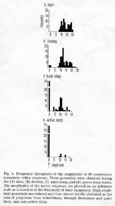
The amplitudes of the reflex motor responses
are plotted in histographic form in Figure 1 for
the 4 states of sleep and wakefulness. During
the alert state a large number of moderate and
high-amplitude reflex potentials - as observed
(Figure 1A). The drowsy state was characerized
by a decrease in the frequency of the
highermplitude potentials (Figure 1B), as
compared with the lert state. During quiet sleep
the mean amplitude of the eflex response
decreased when compared with the drowsy tate
(Figure 1C). At times during quiet sleep reflex
reponses failed to occur following stimulation
of the
mesencephalic nucleus. These events are
indicated by the frequency of zero amplitude
potentials (arrow at zero in figure 1). The
majority of the reflex responses was completely
suppressed during active sleep (Figure 1D).
A statistical analysis of the change in mean
amplitude of the reflex during the successive
states in the sleep cycle utilized planned
comparison tests based on an analysis of
variance. The changes in state - waking compared
with drowsy, drowsy with quiet sleep, and quiet
- sleep with active sleep - were each marked by
a significant reduction in the amplitude of the
reflex response (p<0.05).