Retropharyngeal tendinitis is a rare cause
of intense neck pain and occipital headache. It
is caused by an aseptic inflammatory process in
the longus colli tendon, triggered by deposition
of calcium hydroxyapatite crystal.
Clinically, it can be misdiagnosed as
retropharyngeal abscess, traumatic injury,
infectious spondylitis, cervical artery
dissection, or even meningitis. The diagnosis is
made radiographically by a nearly pathognomonic
amorphous calcification anterior to C1-C2 and
prevertebral soft tissue swelling. We present a
new case of this uncommon condition exhibiting
some unusual features.
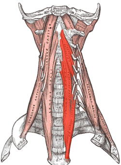
Acute aseptic tendinitis of the longus colli
muscle (retropharyngeal tendinitis) is extremely
rare and was first
described by Hartley
and Fahlgren in 1964. Due to the typically
sub-acute onset of extremely severe neck
pain‚ and more seldom headache‚
painful restriction of movement in the cervical
spine and increased body temperature,
retropharyngeal tendinitis is an important
differential diagnosis in patients with
secondary headaches and neck pain.
CASE REPORT
A 38-year-old female patient was admitted to
hospital following the occurrence of progressive
complaints over the course of only a few days.
She reported continual, sharp and diffuse pain
at the nape of the neck, which radiated up into
the back of the head and was aggravated by head
movement.
The patient also complained of swallowing
difficulties and excoriating pain during
yawning.
No incident of cervical spine trauma was
recalled.
An infection of the upper respiratory tract,
the pharynx or teeth prior to symptom onset was
negated. The patient had been operated on
5-years earlier due to a herniated disc at level
C5/C6. A Bryan cervical disc prosthesis had been
inserted.
Clinical assessment revealed painful
restriction of cervi- cal movement. Neurological
status was normal. An elevated leukocyte count
12.8/nL and CRP 91.3 mg/L was established.
Extensive laboratory analyses, e.g., rheumatoid
factor, ANCA, ANA, revealed normal results.
Normal results were also found in the ENT
examination for the oral cavity, pharynx, and
larynx, ruling out a retropharyngeal
abscess.
Cervical spine x-rays showed that the Bryan
disc prosthesis was correctly positioned and
also revealed kyphosis at C5/C6. Calcification
ventral to C2 inferior to the anterior arch of
the atlas and a widening of the prevertebral
soft tissue shadow was observed. In comparison
with outpatient x-rays taken 1 year earlier were
not present, x-rays were taken in order to check
the position of the inserted Bryan prosthesis),
an increase in the amount of calcification was
evident.
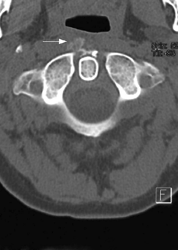
In accordance with the x-ray images,
computer tomography revealed a narrow, band-like
and moderate calcification to the right and
ventral anterior to the first and second
cervical vertebrae. The calcification was
located in the very area of the longus colli
muscle. MRI revealed hypointensity ventral to C2
and a thin prevertebral hyperin- tense rim of
edema. Altogether, the findings resulted in a
diagnosis of acute calcifying prevertebral
tendinitis.
The patient received antiphlogistic
treatment with ibuprofen (500 mg/d t.i.d.). Due
to the severity of pain experienced, initial
treatment with opioids (100 mg tramadolol/d) and
the muscle relaxant benzodiazepine tetrazepam
(50 mg/d) was also necessary. This treatment led
to a rapid alleviation of symptoms, so that it
was possible to discharge the patient only a few
days later.