-
- The act of yawning begins during fetal life.
The complex biochemistry involved in this action
includes many enzymes and neurotransmitters,
including: dopamine, acetylcholine, muscarine,
histamine, adenosine, serotonin, nitric oxid,
adrenocorticotropic hormine, oxytocin,
alpha-melanocyte hormone, opioids and
gammaaminobutyric acid.
-
- An early research into the physiology of
yawning was conducted by Charcot.
His famous case was a woman with relentless
yawning who was hospitalized for months in
Salpetriere hospital. This patient presented
with a yawning frequency of 8 yawns per minute,
and Charcot noted that although her breathing
pattern was severely disturbed her ventilation
was not reduced.
 -
-
- Charcot
then realized that the patient's yawning was
functioning as deep inspirations replacing
normal breathing movements (breathing through
yawning). A single yawn can therefore be viewed
as an isolated act of hyperventilation in which
the oxygen saturation is increased and the PCO2
reduced.
-
- K. P. van de Woestijne and D. Trop have
demonstrated in their research on dogs that
alveoli collapse to 60% of their initial volume
after 2 hours of anaesthesia. A similar finding
has been observed in humans. The alveolar
collapse suggests the existence of shunting of
venous blood causing a decrease in blood
oxygenation. A single deep inspiration or yawn
can restore the alveoli to their initial
capacity. This finding has led Forrester to
suggest that the function of yawning is
maintenance of pulmonary alveolar patency, and
that yawning serves as a defensive mechanism
against alveolar collapse.
- Observations in rats have shown an
association between yawning and erection.
Additional experiments found that castrating
animals causes a decrease in yawning frequency
and that a later course of testosterone
replacement has the reverse effect of increasing
yawning frequency. Since erection is function of
autonomically mediated vasodilation the
connection of yawning to autonomic stimulation
became an area of experimental interest.
-
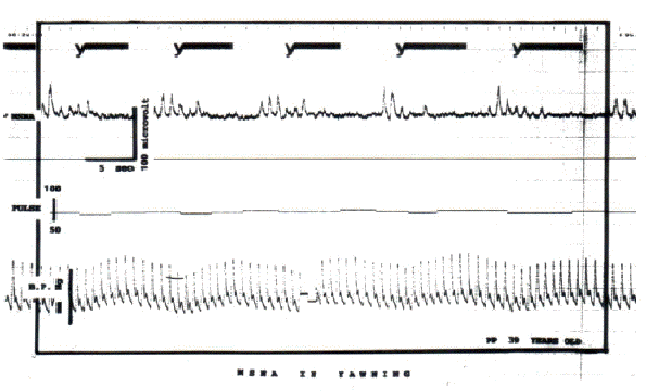
- The deep inspiration produced by yawning
causes the dilatation of lung bronchial muscles,
stimulating a vagal response with discharge of
acetylcholine, and inhibition of
sympathetic&endash;adrenergic activity.
Inhibition of sympathetic activity causes
arterial dilatation which diminishes arterial
resistance and accelerates the arterial
circulation (Friedell 1974, Lehman
1979, Twiest 1974). The decreased sympathetic
activity during yawning was demonstrated by
direct recording with the microneurographic
technique by Askenasy and Askenasy (1996). Shown
in the picture is a decrease in sympathetic
activity following every yawn:
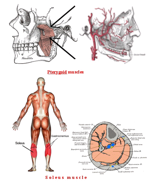
 - During the yawning movement additional
muscular contraction occurs in addition to
diaphragmatic contraction. This movements are
termed gasping (the wide opening of the mouth)
and pandiculation (limb stretch). Gasping and
pandiculation serve a vascular effect by
discharging the venous plexuses located within
the pterygoid and soleus muscles respectively
into the circulation increasing the blood volume
available for oxygenation. This mechanism is
named "the peripheral hearts" or "the sural and
tricipital pump". (Bhangoo 1974,Last 1963).
-
- Arrows point to venous plexus location:
 -
-
- Pterygoid muscles and Soleus
muscle
-
- Yawning is considered by Bell and Suganami
as being provoked by boredom, a consequence of
diminished interest and stimulation by a source
of information. The difficulty in maintaining
cortical attention becomes a neural stimulus
named "down-attention" which announces a change
in the cerebral state.
-
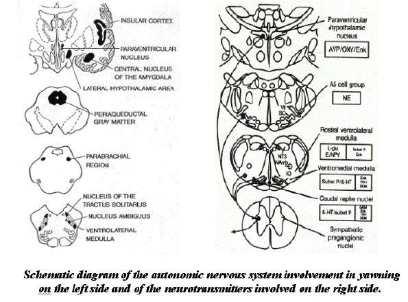
- The harmonic inter-connection between
yawning, breathing and even coughing implies to
the existence of a perfect synchronization
mechanism of the respiratory, cardiovascular,
and muscular systems. Such intricate control is
naturally found in the central nervous system.
The brain areas involved in the elaboration of
such a harmony are: the neocortex, the
límbic system, the
hypothalamic-hypophyseal system and the
reticular system.
-
- Neocortical involvement in the regulation of
yawning is demonstrated by the contagiosity of
yawning. Simply viewing another person yawn may
be a sufficient trigger for a yawn by the
observer (Provine 2005). Involvement in yawn
regulation of the parvocellular oxytocinergic
neurons in the paraventricular nucleus of the
hypothalamic system was proved experimentally by
Kita I et.al, and the involvement of the
autonomic system was proved by microneurography
(Askenasy & Askenasy). Reticular involvement
is the subject of the present presentation.
-
- The generally accepted causes of yawning are
boredom, fatigue and drowsiness. The yawning
center was hypothesized to be localised in the
reticular formation (Askenasy 1989). The
reticular formation is located in the brainstem.
The brain stem is located between the thalamus
and the spinal cord. This structure, with a
width of only an inch has a critical function in
the brain.
-
- The brain relates to the environment through
sensory information reaching the thalamus and to
the body through ascending neurons from the
spinal cord. The Corpus callosum connects the
two hemispheres of the brain, and the pons
(latin for bridge) connects the two sides of the
cerebellum, connecting each side of the
cerebellum with the opposite side and with the
cerebral hemispheres. The Pons also connects the
upper part of the nervous system with the
medulla oblongata. The Medulla oblongata is the
caudal part of the brain stem and sits above the
top end of the spinal cord Both types of inputs
supply the reticular formation. In the time it
takes you to say these two words, the medulla
oblongata will have regulated your breathing,
blood pressure, heart rate and all cathartic
reactions in the states of sleep and
wakefulness.
-
- Salzarulo intuition about the signaling role
of yawning was stipulated in his theory about
the stabilization effect on transitory periods
of the sleep/wake cycle.
-
- Karasawa and his team have studied the
consequences of serial yawning on the
electroencephalography and O2 saturation in
patients with cerebral vascular accidents of the
thrombotic type (1982). They found a significant
decrease in electroencephalographic activity and
yawning associated with a decrease in the
partial pressure of sanguine oxygen, as it
occurs during boredom, somnolence and fatigue.
The authors stipulated that this effect is
caused or provoked by a decreased activity of
the ascending reticular system.
-
- Monatagu
(1962) consider that the reticular system
stimulation is the result of the accelerated
blood circulation and hyperoxygenation.
-
- Robert
Provine, suggested several times yawning as
a result of a disturbed harmony of the nervous
system.
-
- Yawning entails a perfect match of the
rhythmic vital functions of breathing and the
cardiovascular circulation under a harmonic
regulation of the nervous system. The proximity
of the centers of these functions and the
yawning center in the reticular formation
explains the perfect harmony.
-
- Yawning is a signal initiated by the
reticular system and brough to our
consciousness, that a change in the functional
state of the nervous system occurs and need
help.
-
- Any change in the steady state as perceived
by the brain provokes a reaction by the
reticular formation. Therefore a yawn may be
triggered by a diverse array of states which
represent a change in situation: boredom,
sadness, surprise, suffering, fatigue, stress,
somnolence...
-
- The reticular system's anatomy of intense
proximity (under 2 inches) of both activating
and inhibitory neuronal networks may explain why
yawning appears in situation which are
seemingley paradoxal such as boredom and
excitation.
-
- Inaccuracies in the harmonic regulation
function occurs on pathologic basis, at
different levels of the central nervous system,
such as the cortical, limbic, hypothalamic and
autonomic nervous system, but always involving
the essential regulator "the reticular
network".
-
-
-
- References
- Askenasy
JJ., Askenasy N. Inhibition of muscle
sympathetic nerve activity during yawning. Clin
Auton Res 1996. 6:237&endash;239.
- Askenasy
JJM. Is Yawing an Arousal Defense Reflex ? The
Journal of Psychology 1989;123(6):609-621
- Bhangoo, K. S. (1974). Letter. New England
Journal of Medicine, 290, 1440.
- Forester JM. Provocation spinale d'un
réflexe de bâillement. Rev d'oto
neuro ophtalmologie.;23(1): 1951
- Friedell, A. (1974). Letter. New England
Journal of Medicine, 290, 1439-1440.
- Karasawa J, Kuriyama Y, Kuro M, Kikuchi H,
Sawada T, Mitsugi T. 1. Monitoring system of
cerebral blood flow and cerebral metabolism. 2.
Relationship between internal jugular oxygen
tension and cerebral blood flow. Brain-Nerve
34:239-245, 1982
- Last J. Regional and Applied Anatomy (3rd
ed., London.
- Lehmann H.
Yawning : a homeostatic reflexe and its
psychological significance. Bulletin Menninger
Clinical, 43:123-126, 1979
- Montagu
A. On yawning, JAMA, 27, 152, 1962
- Provine
RR (2005). Yawning". American Scientist 93 (6):
532.
- Salzarulo P, Gianluca F. Awakening abd
sleep-wake cycle across development 281 p.,
2002
- Sherwood Lauralee, Fundamentals of
Physiology: A Human Perspective, Thomson
Brooks/Cole, 2006, p. 380
-
-
-
-
|
