- CHYLOTHORAX is a relatively rare condition
characterized by pleural fluid that has a high
concentration of neutral fat and fatty acid but
little cholesterol. Bilateral chylothorax is
even more unusual. Trauma and tumor are
responsible for the majority of the 51 cases of
bilateral chylothorax that we were able to find
in a search of the literature.
-
- Meade has reported five cases of unilateral
chylothorax that he believed were due to
hyperextension of the spine with fixation or
inherent weakness of the thoracic duct. We
describe what we believe is the first reported
case of bilateral chylothorax caused by this
mechanism.
-
- Report of a Case
-
- A 56-year-old woman was admitted to
Georgetown University Hospital on July 9, 1974,
for recurrent pleural effusions.
-
- In October 1973, she had experienced the
sudden onset of sharp, pleuritic chest pain on
the left and swelling above the left clavicle
following an episode of vigorous stretching
while yawning. Chest x-ray film demonstrated
bilateral pleural effusions, greater on the
left. Treatment with antibiotics was followed by
resolution of her symptoms and clearing of the
effusions.
-
- The patient recalled that on the day before
the present admission, a similar episode of
stretching was followed by swelling in the left
supraclavicular fossa, left anterior chest pain,
and a dry, nonproductive cough. Bilateral
pleural effusions were seen on x-ray films.
There was no history of trauma, recent
infection, fever, anorexia, or weight loss.
Medical history included 20 years of vague
arthralgia of the back, knees, and shoulders,
three prior episodes of pneumonia, and a two- to
three-pack/week cigarette habit for 25
years.
-
- Results of physical examination were normal
except for soft-tissue swelling and tenderness
in the left supraclavicular fossa, as well as
flatness of the percussion note and decreased
breath sounds at both lung bases.
-
- The hematocrit reading was 33% and white
blood cell (WBC) count was 5,600/cu mm, with a
normal differential cell count. The Westergren
sedimentation rate was 15 mm/hr. Results of
urinalysis, and values for glucose, blood urea
nitrogen, amylase, electrolytes, calcium,
phosphorous, serum glutamic oxaloacetic
transaminase, serum glutamic pyruvic
transaminase, alkaline phosphatase, and
bilirubin were normal. Lupus erythematosus
preparation, VDRL test, and tests for cold
agglutinins were negative. The fluorescent
antinuclear antibody test was weakly positive.
The complement level was within normal
limits.
-
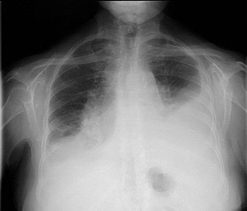
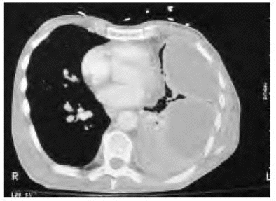
- Initial chest x-ray film disclosed a
moderate amount of fluid in both pleural
cavities. Bilateral thoracentesis yielded
creamy, milky-white fluid. Fluid analysis showed
a red blood cell count of 211/cu mm, a WBC count
of 1,215/cu mm, with 22 polymorphonuclear
leukocytes, 14 lymphocytes, and 64 unidentified
mononuclear cells. The fluid cholesterol level
of 117 mg/100 ml, with a serum cholesterol value
of 171 mg/100 ml, and a positive result on a
preparation stained with Sudan IV for fat,
confirmed the chylous nature of the fluid.
Studies on cells of the fluid and the pleural
biopsy specimen showed no neoplasm, and there
was no growth of bacteria, fungi, or acid-fast
bacilli. Degenerative changes in the cervical,
thoracic, and lumbar regions were seen on x-ray
films. Mediastinal tomograms, intravenous
pyelogram, and scans with gallium citrate Ga 67
were normal. A lung scan did not suggest
pulmonary emboli. Bilateral pedal
lymphangiograms were obtained on the tenth
hospital day, at which time the patient was
asymptomatic with only slight blunting of the
left costophrenic angle on chest x-ray film. The
lymph nodes and thoracic duct were completely
normal.
-
- The patient was discharged asymptomatic to
be observed by her private physician.
-
- Comment
-
- Injury to the thoracic duct below the T-5
level results in a right-sided chylothorax,
while damage to the thoracic duet above T-5
leads to a leftsided chylous effusion, The
thoracic duct usually crosses from the right to
the left side of the thorax at the T3-6 level,
and injury to the vessel at this point would be
expected to result in spillage of chyle into
both pleural cavities. Bilateral chylothorax was
discovered in our patient after a second episode
of bilateral pleural effusion following
stretching. Regrettably, the chylous nature of
the fluid was not confirmed after the first
episode, but the fluid was probably chylous in
the first episode as well as the last since
there was a similarity in signs and symptoms on
admission, subsequent findings, and in the
course of the effusions. We postulate that
sudden hyperextension of the spine during
yawning resulted in injury to a fixed or
inherently weak thoracic duct with leakage of
chyle into both pleural cavities, followed by
healing of the duct and resorption of the
chylous effusions.
-
 
|


