- Tous
les articles consacrés au
bâillement
foetal
- Fetal
yawning: all
publications
-
- Abstract
- The aim of the study was to observe
different expressions and movements of a fetal
face during investigation of fetal behavior in
the second and the third trimester of normal
pregnancies, as a probable manifestation of
fetal awareness.
-
- Over a 6-month period a study was conducted
in three centers in Zagreb, Croatia and in
Barcelona and Malaga, Spain. Women with
singleton pregnancies (16-33 weeks) who were
referred for ultrasound check-up for
determination of gestational age, suspicious
fetal malformations, polyhydramnios, and/or the
assessment of biophysical profile or other
possible pathology, were assigned to the study.
After regular twodimensional (2D) ultrasound
assessment at an antenatal clinic, pregnant
women were offered the possibility of undergoing
4D ultrasound examination if the fetus and the
mother were considered "normal", i.e., if
ultrasound and clinical assessment were
uneventful. If the newborn delivered at term had
1- and 5-min Apgar scores of 7 and 10,
respectively, and if the newborn was considered
"term and normal" (normal spontaneous activity,
normal posture and tone, and presence of some
primitive reflexes) at the first and subsequent
regular check-ups, the inclusion criteria were
deemed to have been met. Out of 119 patients, 99
fulfilled the inclusion criteria, 40 of whom
were in the second, and 59 in the third
trimester of pregnancy. A Voluson 730 Expert
system with a transabdominaI 5-MHz transducer
was used for 4D ultrasonography. After regular
2D scanning, the 4D mode was switched on, and a
live 3D image was reconstructed by selecting
ideal 2D mid-sagittal images of the face (the
region of interest). The volume was
automatically scanned every 2 s while the
surface-rendered mode was switched on, and 4D
images were displayed on the screen and recorded
on videotape during a 30-min observation period.
Movements of the following fetal face structures
were analyzed: forehead, brows, nasal soft
tissue and nasolabial folds, upper lip, oral
cavity and tongue, lower lip and chin, eyelids
and eyes, mouth and mouth angles, and facial
expression. 4D ultrasonography allowed in utero
observations of fetal facial expressions such as
smiling, yawning, and swallowing.
-
- The quality of 4D depiction of fetal facial
expressions increased with gestational age. The
frequency of fetal facial expressions such as
yawning ranged from 1 and 6 with a median of 1.5
per 30-min observation period; smiling ranged
from 2 and 8 with the median of 2; tongue
expulsion ranged from 2 to 6, median 3; mouth
and eye squeezing ranged from 5 to 10, median 6;
scowling ranged from 1 to 3, median 0.5; and
isolated eye blinking ranged from 4 to 12 with a
median of 5.
-
- Our study shows the ability of 4D sonography
to depict different facial expressions and
movements, which might represent fetal
awareness. Nevertheless, long, precise and
thorough observation of fetal faces by 4D
sonography was hampered as the images were only
near real-time. Thus, we were only able to study
the quality and not the quantity of facial
movement patterns.
-
  -
- Introduction
-
- During recent years more has become known
about various components of the development of
the human nervous system and how these
components act in concert during fetal life.
This knowledge has led to a renewed discussion
over whether the developing fetus is capable of
being aware of its state and surroundings and,
if so, when this awareness occurs during
gestation.
-
- For better understanding, it is useful to
consider the fundamental parts of early brain
development. Four critical regions start to
develop from the forebrain after the fifth
gestational week: the thalamus, cerebral cortex,
hypothalamus, and limbic system. The thalamus
becomes the reception area for most of the
sensory input to the brain, which ascends the
spinal cord, and relays it to the appropriate
region of the cortex via its projection fibers.
Thalamocortical fibers begin to develop at 17
weeks and penetrate the cortical plate to make
permanent connections at 22-34 weeks.
-
- The part of the brain associated with
thinking, consciousness, emotions, etc., is the
cerebral cortex, which forms the largest part of
the developed brain, enveloping the lower
structures in two cerebral hemispheres, the
first signs of which are visible at 5-6 weeks.
It has been indicated that facial reflexes in
response to somatic stimuli, which could
indicate an emotional reaction to pain, develop
rather early in gestation. Nevertheless, it is
still questionable whether it is possible to
presume brain function only from fetal behavior.
From the developmental point of view, it is
clear that many facial and encephalic structures
have the same embryologic origin, and therefore
the consideration that "the face predicts the
brain" seems to be true. Whether the fetal face
is an "organ that expresses awareness" with very
complex functions is still questionable, because
we need more scientific confirmation of what
constitutes fetal awareness.
-
- Four-dimensional (4D) ultrasound technology,
introduced in recent years, is a novel tool for
the observation of fetal behavior and the fetal
face. Although two-dimensional (2D)
ultrasonography documents the origin, occurrence
and developmental course of specific fetal
movements, simultaneous imaging of complex
facial movements was impossible using only a 2D
real-time technique. A technique was needed to
enable three-dimensional (3D) imaging of fetal
facial movements in real-time mode. This
technique can be called "live" 3D ultrasound or
4D ultrasound, as coined by a manufacturer,
because time becomes a parameter within the 3D
imaging sequence. Human eyes are able to
differentiate between images with a frequency of
up to 12 images per second; consequently,
production of an appropriate frame rate with
specially designed probes and a fast computer
device is required. At the moment, 4D ultrasound
scanning is not real-time and the machines
available can reach up to approximately 20
images per second, depending on the volume size,
resolution and the mechanics of the probe.
Nevertheless, even at these relatively slow
frame rates the ability to study fetal activity
and superficial structures is remarkably good
compared to 2D ultrasonic devices. This means
that 4D ultrasonography integrates the advantage
of the spatial imaging of fetal structures,
especially the face, with the addition of time,
allowing depiction of the appearance and
measurement of the duration of each movement.
This new diagnostic tool allows continuous
monitoring of the fetal face and other surface
features of the fetus, thus opening exciting new
possibilities for the study of the relatively
unexplored area of fetal facial expressions as a
possible manifestation of fetal awareness.
-
- Is it the facial expression of the fetus
that can help us understand what the fetus would
like to communicate?
-
- As our recent investigation showed, there is
behavioral continuity from fetal to neonatal
life, which probably includes facial
expressions. We can see on the fetal face
whether it is satisfied or unhappy, smiling or
worried, self-confident or uncertain, but does
the expression of the fetal face predict its
normal neurological development ? To many of
these questions there are few answers as
yet.
-
- In our systematic study of fetal behavior by
4D sonography we were able to observe different
expressions and movements of the fetal face, but
the question is whether they indicate fetal
awareness. To the best of our knowledge, the
present study is the first attempt to use 4D
sonography in the evaluation of facial
expressions and facial movements to demonstrate
fetal awareness.
-
- Subjects
-
- Over a period of 6 months (November
2003-April 2004) a study was conducted in three
centers: "Sveti Duh" General Hospital,
Department of Obstetrics and Gynecology, Medical
School University of Zagreb, Croatia (Zagreb);
Department of Obstetrics and Gynecology,
Institut Universitari Dexeus, Barcelona, Spain
(Barcelona); and in Centro Gutenberg, Malaga,
Spain (Malaga). Pregnant women of gestational
age between 16 and 33 weeks and with a singleton
pregnancy, who were referred for ultrasound
examination to a tertiary outpatient clinic due
to undetermined gestational age, suspected fetal
malformations, polyhydramnios, and/or for
assessment of the biophysical profile or other
possible pathology, were recruited for the
investigation. Fetuses before 16 and after 33
weeks of gestation were not excluded, but we had
some technical problems with the visualization
of their faces. The optimal and most
satisfactory visualization of fetal faces was
obtained in the second and third trimesters of
pregnancy. After regular 2D ultrasound
assessment at the antenatal clinic, the pregnant
women were offered the possibility of undergoing
4D ultrasound examination. All participants
provided written informed consent and approval
for the investigation. The local ethical
committees in all centers approved the study.
Patients were offered 4D ultrasonography if both
fetus and mother were considered "normal", i.e.,
if the initial ultrasound and clinical
assessment were uneventful. If the newborn,
eventually delivered at term, had normal 1- and
5-min Apgar scores, was considered "term and
normal" and demonstrated, at the first and
subsequent regular check-ups (at least two),
normal spontaneous activity, normal posture and
tone, and the presence of some primitive
reflexes, then the inclusion criteria were
deemed to have been met. Out of 119 patients, 99
fulfilled the inclusion criteria, 40 of whom
were in the second and 59 in the third trimester
of pregnancy.
-
- Methods
-
- 4D ultrasound : All 4D examinations were
carried out using a Voluson 730 Expert system
(GE Medical Systems, Milwaukee, WI, USA and
Solingen, Germany) using a transabdominal 5-MHz
transducer. After standard assessment using 2D
B-mode ultrasound, the 4D mode was switched on
and "live" 3D images were reconstructed by
selecting an ideal 2D mid-sagittal image of the
face (the region of interest; ROI). The crystal
array of the transducer swept mechanically over
the ROI. The volume was automatically scanned
every 2 s while the surface-rendered mode was
switched on, and 4D images were displayed on the
screen and recorded on videotape during a 30-min
observation period. This procedure was used for
the observation of fetal face movements and
expression. From top to bottom, the following
landmarks were analyzed: forehead, brows, nasal
soft tissue and nasolabial folds, upper lip,
oral cavity and tongue, lower lip and chin,
eyelids and eyes, mouth and mouth angles, and
facial expression. All recordings were performed
between 14.00 and 17.30 h, and no meal was taken
within the 2-h period of the study.
-
- Definitions
-
- Awareness was defined as the ability to
notice something using senses - a phenomenon
incorporating both cognitive and physiological
elements. There is urgent need for more
scientific confirmation of fetal awareness,
starting from its definition through to
reproducible evaluation. Very early in pregnancy
fetuses are reactive to stimuli, but the
reaction does not provide any evidence that the
fetus actually experienced the stimulus. It has
been shown that noxious stimuli can initiate
physiological, hormonal, and metabolic
responses, but these neither imply nor preclude
suffering, pain, or awareness.
-
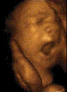
- 4D ultrasonography allowed in utero
observations of fetal facial expressions of
smiling, yawning, and swallowing.
-
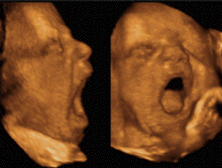
- Yawning
consists of breathing through the mouth and
nose, whereby a long inhalation with the mouth
wide open is followed by a slow exhalation. A
single, continuous opening of the mouth can last
for 4-6 s. The anatomical criterion for fetal
yawning is retraction of the tongue, whereas
yawning in adults is characterized by an
extended tongue. Yawning develops at 11.5-15.5
weeks' gestation.
-
- The three phases of swallowing (oral,
pharyngeal, and esophageal) are the same during
fetal life and afterwards. The oral and
pharyngeal phases appear to be less well
developed in fetuses. In a mature fetus, two-six
sucking movements usually precede the initiation
of the oral stage of swallowing; thus, the
swallowing pattern in a normal fetus near term
differs from that in the infant and in the
adult. The human fetus demonstrates swallowing
movements as early as 11-12.5 weeks' gestation,
whereas more complex sucking movements can be
identified at 18-20 weeks. Neurobiological
control of fetal swallowing includes coordinated
contractions of the thyroid, nuchal, and
thoracic segments of the esophagus. Although
swallowing may bring satisfaction to the fetus,
it is very difficult to correlate 4D sonography
with a possible indication of fetal
awareness.
-
- Scowling, smiling, isolated eye-blinking,
tongue expulsion, and mouth and eye squeezing
are obvious facial expressions or activities
that can also be observed by 4D sonography.
-
- Although there are reports in the literature
that the "quality" and not the "quantity" of
general movements in neonates is a better
predictor of neurological outcome, the quality
of facial movements has been neither described
nor studied. Positive observation has been
defined as a facial expression or movement noted
at least once during the observation of one
examinee.
-
- Results
-
-
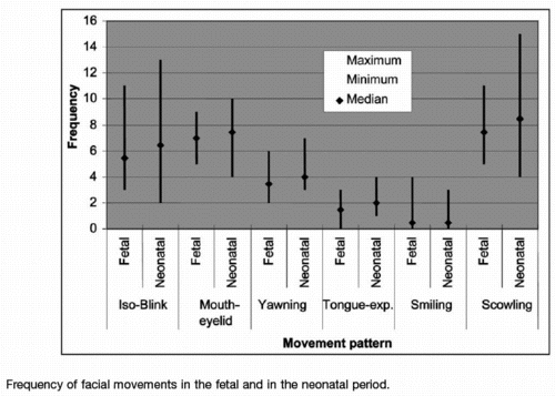
- We noted a tendency towards increased
frequency of observed facial expressions with
increasing gestational age, but the difference
between second- and third-trimester fetuses was
not significant due to the low frequency of
movements. Therefore, all observed facial
expressions and movements (i.e. yawning,
smiling, tongue expulsion, mouth and eye
squeezing, scowling, and isolated eye blinking)
are presented collectively in Figure 2, with
maximum, minimum, and median frequencies during
a 30-min observation period. Some fetal facial
expressions are shown in Figures.
-
- Discussion
-
- Ultrasound is one of the most rapidly
developing medical technologies that can extend
the physician's inspection, palpation and
auscultation. Ultrasound has been very important
for the developmental assessment of the fetal
central nervous system and its behavioral
patterns throughout gestation , and it is
generally accepted that patterns of fetal
activity reflect the development and maturation
of the central nervous system.
-
- We are aware that some postnatal studies
distinguish between "quantity" and "quality" of
motor patterns. Quantity is the number of fetal
movements expressed as a percentage of
observation time or as the number of events per
time epoch. The quality of each individual
movement includes speed, amplitude, and force
combined in one complex perception. It has been
suggested that the observation of behavioral
quality is a better predictor of neurological
impairment than neurological examination. In
this respect, we are unable to study the quality
of facial movements in fetuses, because this
parameter has not been described yet.
-
- Although the advancement of technology
enables depiction of the fetal face in real time
and observation of its facial expression, there
are still many improvements to be made. Since 4D
ultrasonography provides only a near real-time
reconstruction of the fetal face, some very
subtle facial movements may have been missed. To
avoid this obstacle, faster frame rates should
be achieved. A prolonged observation period
could possibly determine if the fetus being
observed is either quietly or actively sleeping,
drowsy, or alert and inactive. Otherwise, the
fetal face will frequently be missed and be
unavailable for proper observation.
-
- 4D sonography, introduced in recent years,
is a completely new tool for observing fetal
behavior and the fetal face. However, as for any
other new technology, there is much room for
improvement. Furthermore, accurate and reliable
fetal face evaluation is still time-consuming.
However, this is the only available method
enabling continuous monitoring of the fetal face
and other surface features of the fetus - facial
expressions - as possible measures of fetal
awareness. Our study shows the possible
visualization of several facial expressions, but
it is still unknown if fetal face expressions
can help us in understanding what the fetus
would like to communicate.
-
- Yawning can
occasionally be observed during the first and
second half of a normal pregnancy. It has also
been shown that term human fetuses yawn
predominantly during active sleep, but not
during quiet sleep. Sherer and associates
described in detail a single yawning movement in
a 20-week fetus; they observed that the fetal
mouth, previously closed, opened widely and
remained so for 2-3 min in association with
simultaneous extension of the fetal arms and
flexion of the fetal head. The physiological
role of yawning during intrauterine life remains
speculative. A respiratory function is less
probable, since thp surrounding environment is
liquid rather than air. However, since a forced
inspirium is a critical component of yawning, a
potential role for expanding the terminal
alveoli in the fetal lungs by the inspired fluid
is possible, consistent with the hypothesis that
yawning may serve as a mechanism to protect
against alveolar collapse in extrauterine
life.
-
- Far too many interpretations of an open
mouth as yawning have been described in
the recent literature. The range of variation
includes, for example, a single continuous
opening of the mouth for 4-6 s. There is also
controversy about the use of the anatomical
criterion of tongue retraction to characterize
the fetal yawn. It seems that there is no
physiological reason why the fetus should yawn.
Doppler studies found no connection to hypoxia.
Furthermore, yawning confers no protection
against atelectasis in a fluid-filled lung. With
2D sonograpic visualization of fetal yawning,
one of the most uncommon behavioral states in
human fetuses could be incidentally observed
during ultrasonic examination of the fetal face.
Yawning is a poorly understood complex movement
present in almost all mammals. Although it can
be induced by several physiological processes,
such as hunger, fatigue, sleepiness, boredom,
and drowsiness, a number of neurological
disorders and drugs can also trigger this
reflex. The physiological function and
neuroanatomical pathways involved in yawning are
still unknown. It is presumed that yawning in
humans can be used as an index of dopaminergic
system functioning.
-
- A detailed study of the normal development
of swallowing over the course of fetal life may
lead to early identification of potential
neonatal swallowing difficulties and may clarify
the mechanism of polyhydramnios and
oligohydramnios. The normal pharyngeal stage of
swallowing in adults includes glottal closure to
prevent aspiration, and opening of the
esophageal sphincter. In the mature fetus, the
trachea is not sealed completely by glottal
closure, and any amniotic fluid swallowed is
directed not only into the esophagus, but into
the trachea as well. According to our
observations, the frequency of swallowing
movements was the same, irrespective of
gestational age.
-
- The introduction of 3D/4D sonography is a
turning point in fetal facial examination. Once
the midsagittal plane is obtained, the volume
dataset can be acquired. First, the
surface-rendering mode is used to search for
facial dysmorphogenesis. Then the multiplanar
re-slicing mode is used, and the three reference
planes, sagittal, axial and coronal, are
simultaneously displayed and scrutinized. Up to
now, examination of the fetal face was only an
integral part of fetal ultrasound examination
during pregnancy, whether in a screening setting
or during targeted analysis. Our study is the
first attempt to use 4D sonography in the
evaluation of fetal facial expressions and
facial movements in order to observe fetal
awareness. Postnatal investigation of facial
expressions of premature infants will help us in
choosing prenatal observations of fetal facial
movements.
-
- Conclusion
-
- Our study shows the ability of 4D sonography
to depict different facial expressions and
movements, which might represent fetal
awareness. Nevertheless, long, precise and
thorough observation of the fetal face by 4D
sonography is hampered by the only
near-real-time images. Development of the
technology will allow even better depiction of
fetal facial expressions and movements. At
present, fetal facial expressions and movements
depicted by 4D sonography can be used as a sign
of fetal awareness. There is an urgent need for
further studies in order to compare the
existence of fetal awareness by 4D
ultrasonography in normal and abnormal fetuses,
which might be helpful in the evaluation of
fetal neurological status. In the meantime, the
new technique should be neither overestimated
nor underestimated.
-
 -
|


