- I enjoyed reading Dr Goodman's clinical
memorandum on fractures of an ossified
stylohyoid ligament in the february Archives
Otolaryngol 1981; 107; 129-130
-
- I recently had a case that illustrates some
of his points:
-
- Report of a Case : A 52-year-old man
with diabetes, previously disabled by a low-back
injury, consulted me about a lump and soreness
in the right submaxillary area of three weeks'
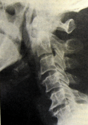
duration. Physical examination disclosed a bony,
hard mass in the submaxillary triangle separate
from the mandible and fixed. Roentgenographic
examination showed an unusually large and
well-developed stylohyoid bone. This measured
about 7 cm in length, with the maximum width of
the shaft about 9 mm. There was a well-defined
medullary cavity and bony cortex, an
articulation proximally at the styloid process,
and a somewhat less well-defined apparent
articulation distally at the anterolateral
aspect of the hyoid bone.
-
- Two days later, while the patient was
yawning vigorously, he heard a loud
crack. Acute pain developed in the area. The
following day, a roentgenogram showed a fracture
with slight angulation in the middle portion of
the shaft of this stylohyoid bone.
-
- Comment : We elected to treat this
surgically by removing the distal portion
through an external approach. The patient had an
uneventful postoperative course and has since
then been asymptomatic.
-
 -
Fracture
of an ossified stylohyoid
ligament
- Archives Otolaryngol
1981;107; 129-130
-
- The styloid process of the temporal bone
varies in length frorn a long bone, reaching
almost to the hyoid and palpable in the
tonsillar fossa, to a tiny structure barely
visible in the dried skull, and difficult to
identify roentgenographically. It is connected
to the hyoid bone by the stylohyoid ligament,
which may be ossified to a variable degree.
Anatomic variations of the styloid bone and
stylohyoid ligament may present confusing
clinical and roentgenographic pictures.
-
- Report of a Case : A 51-year-old
woman was in good health until she lost control
of her automobile at low speed and drove it into
a tree. She was thrown forward by the impact and
struck the steering wheel with her neck. When
brought to the emergency room, she was in no
respiratory distress but complained of neck pain
and hoarseness. Lateral neck films were obtained
and initially seemed to show a fracture of the
hyoid bone with soft-tissue swelling and airway
compromise. I was called to see the patient
immediately.
-
- On examination, the patient was
uncomfortable but in no acute distress. There
was no stridor. Her neck was diffusely tender,
but all landmarks were palpable and intact.
Indirect laryngoscopy showed the airway to be
widely patent. The vocal cords wee mobile, and
there were no lacerations or hematomas of the
laryngeal mucosa.
-
- The patient was hospitalized for observation
and given 100 mg of methylprednisolone sodium
succinate (Solumedrol) intravenously to prevent
tissue swelling. A tracheotomy set was placed at
her bedside, and she was informed that an
emergency tracheotomy might be necessary in the
event of tissue swelling and airway compromise.
During the next 48 hours, she had no respiratory
difficulty, and repeated examination results
were normal. The neck pain and general
tenderness gradually subsided; the tenderness
could then be localized to the left side. At the
time of her discharge from the hospital, there
were still pain and a grating sensation in the
left side of the neck on swallowing. An
anteroposterior view of the neck demonstrated
that an ossified stylohyoid ligament was present
in the left side of her neck.
-
- Comment : The styloid process and the
lesser cornu of the hyoid bone develop from the
cartilage of the second branchial arch.
Connecting them is the stylohyoid ligament. In
some mammals it is normal to find an ossified
chain in place of this ligament; in humans this
is an uncommon (but not rare) anatomic variant.
In one series, ossification of the stylohyoid
ligament was found in 23 of 516 unselected neck
films, in patients as young as 19 years. It has
been noted in patients as young as 2 years. This
strongly suggests that the cause is not
degeneration with calcification, but true
ossification.
-
- In the human embryo, the epihyal cartilage
is resorbed, and its fibrous sheath persists as
the stylohyoid ligament. Failure of this step
leaves cartilage that may ossify. Instead of a
single structure, there may be a chain of three
or four bony parts, reflecting the multiple
cartilages of the embryo. The width of the
structures, length, and the degree of
ossification are highly variable. Ossification,
when it occurs, is usually (but not always)
bilateral.
-
- Because it is uncommon, an ossified
stylohyoid ligament may prove confusing.
Roentgenographically it bas been mistaken for a
foreign body, with some patients undergoing
endoscopies because of it. On palpation it may
mimic a tumor. In this case the roentgenograms
were misread as showing a fracture of the hyoid
bone. Porrath, states that hyoid fractures are
unusual even in cases of direct trauma. This is
because of the mobility of the bone and its
protection by a covering of soft tissue.
However, he believes that hyoid fractures may
accompany mandibular fractures more commonly
than is appreciated. Papavasiliou and Speas on
the other hand, argue that hyoid fractures may
only seem to be unusual because such an injury
often causes asphyxiation before the victim can
be examined. They observed two patients with
hyoid fractures, both of whom required
tracheotomies. In one of these patients the neek
appeared normal after the injury. However,
Porrath states that fracture of the hyoid bone
does not necessarily indicate laryngeal damage
and that, in the absence of other findings,
conservative treatment may be appropriate unless
there is laceration of the mucosa.
-
- It is impossible to generalize about
fractures of an ossified stylohyoid ligament, as
this has apparently been reported only once in
the past. In that case, the patient complained
of a persistent foreign-body sensation. The
patient may have sustained the fracture through
muscular action while retching on a piece of
meat, as violent muscular contractions have been
reported to cause hyoid fractures.
-
- An elongated styloid process can cause vague
discomfort in the throat or even pain (Eagle's
syndrome). The problem in these cases is often
elusive but can be determined by palpating the
bone in the tonsillar fossa or by
roentgenography. The syndrome has been treated
by transoral excision of the tip of the bone,
but also by deliberate fracture. No ill effects
have been reported in these fractures, and it is
likely that a fracture of an ossified stylohyoid
ligament would be similarly benign. It does,
however, serve as an indicator of neek trauma
and require further investigation.
-
- Roy S. GOODMAN, MD
- Barstow Medical Center
- 29 Barstow Rd
- Great Neck, NY 11021
-
|


