- Introduction
Obstructive sleep apnoea (OSA) is a serious
breathing problem that affects approximately 4%
of adults. OSA is associated with increased risk
for adverse cardiovascular events such as
angina, myocardial infarction, stroke and
daytime hypertension. It also has adverse
effects on sleep regulation, producing excessive
daytime sleepiness, impaired work performance
and increased risk for vehicular accidents, and
impaired ventilatory and arousal responses to
hypoxia and hypercapnia. Overall, OSA is a
significant public health problem, with adverse
clinical, social and economic consequences.
Current treatments
A detailed critique and comparison of current
treatments for OSA is outside the scope of the
present review, but both surgical and
nonsurgical approaches (e.g. continuous positive
airway pressure [CPAP], oral appliances
and weight loss) all have some success in
reducing the severity of OSA. With the exception
of CPAP, however, no current treatment is able
to abolish apnoea effectively across all sleep
states, and some treatments have only minimal
effects. Nevertheless, although CPAP at
appropriate pressure is effective in abolishing
apnoea, patient compliance is a serious problem
and impaired daytime function returns after
missing only one night of treatment.
Sleep mechanisms are critical to
obstructive sleep apnoea
Pharyngeal muscle tone Suppression of
pharyngeal muscle activity in sleep is critical
to OSA by producing a narrower airspace that is
more vulnerable to collapse on inspiration.
Anatomical factors that result in a narrowed
upper airspace (e.g.pharyngeal fat deposition,
hypertrophied adenoids and tonsils,
retrognathia, micrognathia, macroglossia)
predispose to OSA by reducing the critical
pressure that is needed for suction collapse.
Likewise, changes in respiratory control system
stability and decreased lung volume in sleep may
also play a role in OSA. Notwithstanding the
importance of such factors in predisposing to
OSA, it is important to emphasize that,
regardless of the features an individual patient
may have that predispose to OSA, the upper
airway still remains open in wakefulness and
closes only in sleep. This simplistic, yet
important, observation highlights a crucial
feature relevant to this review, namely that OSA
is a disorder dependent on sleep mechanisms
because occlusions occur only in sleep. By
extension, even in individuals with structural
narrowing of the upper airway, OSA is ultimately
caused by the impact of brain sleep mechanisms
on the processes that control motor outflow to
the pharyngeal muscles, the tone of which is
necessary and sufficient to keep the airspace
open during wakefulness.
Reflexes
The asphyxic stimuli and suction pressures
generated during airway obstruction in sleep do
not activate the pharyngeal muscles sufficiently
to relieve the obstruction if the patient does
not arouse from sleep, further highlighting the
significant role of sleep mechanisms in OSA.
Importantly, OSA patients also exhibit increased
genioglossus (GG) muscle activity during
wakefulness, suggesting the presence of a
neuromuscular compensatory mechanism that
prevents upper airway collapse in those
individuals with narrowed airways. Although the
mechanisms producing this compensatory increase
in pharyngeal muscle activity in OSA patients
are unknown, it is significant that this
compensatory reflex is present in wakefulness
and its withdrawal in sleep precipitates
OSA.
Summary
In order to understand the pathogenesis of
OSA, it is important to identify the
mechanism(s) that underlie the wakefulness
stimulus' to the pharyngeal dilator muscles.
Specifically, it is necessary to identify the
neurochemical basis of the effects of sleep and
wakefulness on both pharyngeal muscle tone and
reflex responses, and especially the mechanisms
that underlie the sleep-dependent loss of the
neuromuscular compensation for the narrowed
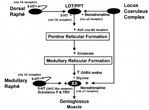
airspace (Fig. 1). Identifying the neural
substrate(s) for the wakefulness stimulus for
pharyngeal motor neurones, and preventing loss
of this stimulus in sleep, may theoretically
lead to prevention of the critical reduction in
pharyngeal dilator muscle activity that
ultimately precipitates OSA. The following text
summarizes some of the brainstem mechanisms that
may be involved in modulating pharyngeal muscle
activity during sleep and awake states, and that
may represent potential therapeutic targets in
OSA. The discussion does not focus on the
general field of pharmacological interventions
in OSA (e.g. use of protriptyline, progesterone,
theophylline, acetazolamide; for overview see),
but for the reasons discussed above it is
restricted to influences of sleep-state
dependent neural systems.
 - [...] Conclusion
There have been several previous attempts in
humans to increase upper airway muscle tone and
to alleviate obstructive apnoeas by
neurochemical approaches, and a resurgence of
interest in these approaches has occurred as
knowledge of the neural systems that affect
pharyngeal motor control increases. To date,
however, these clinical studies have met with
only limited success, in large part because the
basic mechanisms that underlie suppression of
upper airway muscle activity in natural sleep,
and the neurotransmitters and receptor subtypes
that are importantly involved, have not yet been
fully determined. Once these neural systems and
receptors have been identified and their
relative importance determined, however, it is
expected that more rational and systematic
approaches can be devised for the systemic
administration of drugs in order to centrally
modulate motor output to the pharyngeal muscles.
Indeed, as in other disciplines (e.g. the
continuing development of drugs for asthma,
heart disease, etc.), an effective route for
overcoming the many obstacles in this field will
probably be forthcoming, especially after the
basic physiological experiments guide the
clinical and therapeutic approaches to target
specific receptors.
From a clinical perspective, the importance
of understanding basic neural mechanisms of
pharyngeal motor control, especially the
differences in neurobiology between non- REM and
REM sleep, cannot be emphasized enough, both in
adequate interpretation of clinical data and in
planning therapeutic interventions. For example,
if progressive inhibition or absence of
facilitation significantly contributes to
further GG muscle suppression from non-REM to
REM sleep, then a suitable combination of
neuropharmacological agents may be more
beneficial to maintaining pharyngeal muscle tone
in REM sleep than modulating a single
neurotransmitter that may only be effective in
non-REM sleep. The implication of this
consideration is that any potential therapy may
have to be tailored to the individual patient,
based on whether their sleep-disordered
breathing predominates in non-REM and/or REM
sleep. Accordingly, all studies investigating
potential treatments for sleep-disordered
breathing should rigorously control for such
variables that influence OSA, such as sleep
stage and even body position in which apnoeas
occur.
|

 .
.