We report two cases of brainstem stroke
involving the upper pons and the
ponto-mesencephalic junction, presenting with
transient excessive pathological yawning,
associated with gait ataxia and in one subject
by upper limb and facial hemiparesis. In these
patients we hypothesise a causal relationship
between the brainstem lesion and pathological
yawning, possibly related to denervation
hypersensitivity of a putative brainstem centre
of yawn. Excessive yawning can be a heralding
sign of brainstem ischemia.
INTRODUCTION
Yawning is a very common and
phylogenetically old behavioural event that
occurs in vertebrates under different
conditions. A yawn consists of a stereotyped
behavioural pattern that begins with an
inspiration associated with marked dilatation of
the pharynx. At the peak of inspiration there
are associated facial movements and the final
part of yawning is passive rapid expiration.
During yawning a coordinated sequence of events
takes place, involving facial, oropharingeal,
tongue, and respiratory muscles, associated with
activity in the axial extensor and limb extensor
muscles and with autonomic changes characterized
by an increased parasympathetic
outflow.[1] The physiological stimuli
that give rise to the yawning response and its
functional significance are not clear. It has
been shown that yawning frequency is not
modified by hypercapnia or by pure oxygen
breathing, it does not seem therefore to have a
straightforward respiratory function.[2]
Yawning occurs preferentially in conditions of
low vigilance and causes transient increases in
arousal as indicated by EEG desynchronisation
though an active role in the maintenance of
arousal has not been demonstrated. The social
importance of yawning is particularly evident in
mammals, where it seems to have a communicative
role in conditions of decreased
vigilance.[2] The neural structures that
control yawning are presumably located in the
brainstem near or within other respiratory and
vasomotor centres, especially those that control
facial mimics, mastication, throat and
respiration and possibly stretching.[3]
Excessive or pathological yawning, is defined as
a compulsive, repetitive action that is not
triggered by appropriate stimuli such as fatigue
or boredom. We describe here two cases of
excessive yawning behaviour associated with
ischemic lesions in the brainstem.
CASE REPORTS
Patient 1: A 74 years old male was admitted
to our clinic complaining of unsteadiness of
stance and gait lasting for 12 hours. The
patient referred the acute onset of excessive
repetitive, compulsive yawning that he was
unable to control; the yawns were repeated at a
frequency of about 3 per minute. Forty minutes
later the patient noticed also gait ataxia and
inability to stand without assistance. When
admitted to the hospital, the neurological
examination showed a slight intention tremor of
the left arm and slight dysmetria in the
finger-to-nose manoeuvre; no limb weakness was
present and tendon reflexes were normal. The
patient was able to stand and walk but the gait
was possible only with enlarged base, irregular
steps and leftward veering. Cranial nerves were
unaffected and nystagmus was not present. The
state of vigilance was constantly normal. The
patient reported abnormally frequent yawning for
three days following the acute onset with
progressively longer intervals between one
yawning act and the other. Three days later the
neurological examination was normal and all
symptoms had disappeared. An MRI scan, executed
3 days after the onset of the neurological
deficit, showed a small hyperintense lesion in
the left paramedian region of the middle pons on
fluid attenuated inversion recovery (FLAIR)
images (Figure 1). The lesion was also evident
as an area of hyperintense signal in T2-weighted
images. At a three months follow-up the patient
was free of all symptoms.
Patient 2: A 66 years old woman presented
with acute-onset unbalance of stance and gait,
followed two hours later by a single episode of
vomiting and by weakness of the left upper limb.
She reported that the symptoms were preceded 20
to 30 minutes before by unjustified excessive
yawning, at a frequency of approximately one
event every 2 minutes. On admission to hospital
the clinical examination disclosed in the
cranial district a slight left lower facial
paresis, a horizontal nystagmus beating
leftwards and a right-sided internuclear
ophtalmoplegia. A pronator drift in
antigravitary posture and clumsiness in distal
finger movements were observed in the left upper
limb. Slight proximal weakness was present also
in the left lower limb. Tendon reflexes were
normally elicitable in the four limbs. An
extensor plantar response was present on the
left side. Finger to nose and reaching
manoeuvres showed slight dysmetria on the right
and could not be evaluated on the left due to
the motor deficit. No sensory deficit could be
observed both in the trigeminal and somatic
territory. The patient showed wide-base gait and
a marked left lateropulsion on stance. Vigilance
was normal. An MRI scan obtained at 5 days from
the onset of symptoms showed a right pontine
ischemia (Figure 1) and MRI-angiography
disclosed a pseudoocclusive stenosis of the
basilar artery. The frequency of yawning
gradually decreased and returned to normal
within 36 hours. The motor deficit on the left
side and the gait ataxia was still present,
though moderately improved, at three weeks from
onset.
DISCUSSION
We describe here for the first time two
patients with brainstem ischemic stroke
presenting with excessive yawning. The possible
causal relationship between the brainstem lesion
and the excessive yawning behaviour could
provide useful information on the anatomical
location of the neural systems controlling
yawning in humans.
The central anatomical pathways subserving
yawning have not been clearly
defined.[4] The evidence in literature
indicates the presence in mammals of a
sub-cortical circuit mediating the yawning
phenomenon, involving the hypothalamus, the
midbrain and the reticular formation of the pons
and medulla.[2][3][4] In
the rat experimentally induced excessive yawning
behaviour can be produced by direct or indirect
activation of the oxytocinergic neurones in the
paraventricular hypothalamic nucleus, which is
thought to play a primary role in initiating the
yawning phenomenon. The activity of hypothalamic
yawning related neurons undergoes a complex
pharmacological control, being enhanced by
dopamine D2 and possibly D3 agonists, nytric
oxide, acetylcoline and ACTH Ð MSH peptides,
orexins and serotonin and downregulated by
opioids.[5] Similar pharmacological
mechanisms may act in humans, where D2 agonists,
SSRI agents and withdrawal from morphine exert a
facilitatory effect on the yawning behaviour.
Also Valproate overdose, Imipramine and
oestrogen substitution may cause pathological
excessive yawning.[5]
The existence also in humans of a putative
yawning centre in the lower brainstem is
suggested by lesional data. Three
reports[6][7][8] have
described patients with locked-in syndrome, with
preserved yawning movements and complete
volitional paralysis of the bulbar musculature.
Also, it has been observed that yawning
movements persist in anencephalic
infants.[2] Up to now the existence of a
cortical representation of yawning has not been
clearly demonstrated, though a recent
brain-imaging work demonstrated the presence of
an area in the posterior cingulated cortex that
is activated by observation of yawning and is
supposed to be involved in the well-known
phenomenon of contagious yawning.[9] In
both our cases, we observed excessive yawning
behaviour associated with a brainstem
infarction. The lesion was located in the
paramedian region in the ponto-mesencephalic
junction in both patients, though the lesion in
patient 2 was much more extended caudally,
involving also the upper half of the pons
(Figure 1). The clinical picture was
characterised by gait ataxia in both patients
which is known to occur extremely frequently in
paramedian mesencephalic and pontine
infarction.[10] [11] Only in
patient 2, due to the anterior extension of the
lesion also a motor deficit was present. Focal
brainstem lesions have already been reported to
cause pathological yawning.
Jurko et al.[12] reported excessive
yawning during hyperventilation in patients who
had previously undergone thalamotomy or with
recent head trauma and concluded that excessive
yawning can be a sign of brainstem damage. None
of our patients did report a facilitatory effect
of hyperventilation.
Arai et al.[13] reported excessive
yawning in a patient with tumour of the floor of
the fourth ventricle and Postert reported
excessive yawning as a symptom of brainstem
localization of multiple sclerosis.[14]
Additionally, excessive yawning has been
observed in progressive supranuclear palsy,
intracranial hypertension and in temporal lobe
epilepsy, though it was not given a specific
value in the localization of the epileptic
focus.[15] [16] The exact
mechanism of excessive yawning following focal
brain lesions is not fully understood. Possibly
the pathological behaviour is the expression of
the liberation from the control of more cranial
structures of a putative yawning centre, caudal
to the lesion, analogously to the hypothesis
postulated for hiccups caused by medullary
lesions[17] or for the symptom of
excessive yawning behaviour in patients with
ALS.[18] Also in our two patients we
hypothesise that the pathogenesis of the
excessive yawning could be related to a
denervation hypersensitivity mechanism. To our
knowledge, this is the first report of excessive
yawning after brainstem stroke and, more
importantly, in both patients yawning appeared
as the earliest symptom reported of the ischemic
insult. We conclude that excessive yawning can
be a presenting symptom of an acute brainstem
lesion and should not be overlooked.
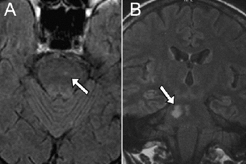
Figure 1: Fluid attenuated inversion
recovery (FLAIR) brain images of the two
patients. A) Patient 1. Axial section
showing a small hyperintense left paramedian
area at the ponto-mesencephalic border. The scan
was acquired at 3 days
from onset of the symptoms B) Patient 2.
Coronal section, showing the ischemic area in
the right paramedian pons and
ponto-mesencephalic border. The scan was
acquired at 5 days from onset of the
symptoms
REFERENCES
Askenasy
JJ, Askenasy N. Inhibition of muscle
sympathetic nerve activity during yawning. Clin
Auton Res 1996;6(4):237-9.
Sandyk
R. Excessive yawning and progressive
supranuclear palsy. Int J Neurosci
1987;34(12):123-4.
Park MH, Kim BJ, Koh SB, Park MK, Park KW,
Lee DH. Lesional location of lateral medullary
infarction presenting hiccups (singultus). J
Neurol Neurosurg Psychiatry 2005;76(1):958.
Williams DR. The
yawning reflex: an upper motor neuron sign in
amyotrophic lateral sclerosis. Neurology
2000;55(10):1592-3.
Response
(O. Walusinski)
Yawning :
a behavior testifying arousal reinforcement
during brainstem stroke.
The two case reports by Cataneo et al. have
the great interest to complete the knowledge
about yawning. Our purpose is to give another
view of the meaning of excessive yawning
observed during brainstem stroke. Among mammals,
there are three types of morphologically
identical yawns occurring in three distinct
situations: situations relative to circadian
rest-activity rhythms, situations relative to
feeding, situations relative to sexuality or
social interactions (1). Until now, no specific
cerebral structure has been identified as a
yawning centre.
A good number of clinical and
pharmacological arguments indicate that yawning
involves the hypothalamus (particularly the
paraventricular nucleus), the bulbus and pontic
regions, with frontal region connections in
primates and to the cervical medulla (2). During
the few hours of life of anencephalous babies,
it has been noted that they yawn and stretch, a
sign of the mammalian syndrome of awakening
activity or « Rekel Syndrom » (3).
Patients afflicted with the locked-in syndrome,
still yawn, despite being paralysed (4,5,6,7).
This shows that yawning originates in the
brain's archaic structures common to all
vertebrates. The central nervous system is based
on a common overall organisational plan and
reveals, from the most ancient to the most
recent vertebrates, a gradual increase in
complexity corresponding to life levels that are
increasingly independent and functionally
developed. Yawning helps understand the
phylogenesis of the encephalon by inferring a
functional organisational pattern of the nervous
system similar to that advanced by Paul MacLean
(8) with the superposition of: (a) an ancestral
"reptilian" brain (brain stem and diencephalon),
where yawning originates; (b) a "paleomammalian"
brain (limbic system) common to all mammifers,
functioning as a synaptic and humoral interface,
in fact the seat of the monkeys' emotivity yawn;
(c) a "neomammalian" brain characterised by
human's cortical development, particularly the
frontal lobes, seat of the "contagious"
yawn.
The phylogenetic appearance of sleep
proposes that the nocturnal rest of
poikilotherms most probably becomes in mammals a
form of the rapid eye movement sleep (REM sleep)
or paradoxical sleep which is caracterized by
peripheral muscular atonia originating in the
dorsal part of the brainstem, rostral to the
pons (9). The ultrasound investigation specifies
is ontogenesis precociousness between 12 and 15
weeks of gestation. Based on numerous studies of
fetuses and infants in a variety of mammalian
species, it is widely believed that the earliest
form of sleep is properly characterized as
active sleep, that is an immature form of REM
sleep and preponderant at birth. Accordingly, it
is thought that quiet sleep, an immature form of
slow-wave sleep (SWS), emerges as REM sleep's
predominance diminishes during ontogeny.
Behavioral pattern continuity from prenatal to
postnatal life shows a strict parallelism
between the ontogeny of REM sleep and yawning.
Basically, REM sleep in the human declines from
50% of total sleep time (8 h) and a frequency of
30 to 50 yawns per day, in the fetus and
newborn, to 15% of total sleep time (1 h) and
less than 20 yawns per day, in the adult. This
decrease takes place mainly between birth and
the end of puberty. As a flip-flop switch, the
reciprocal interactions between sleep and wake
promoting brain regions allow the emergence of
distinct states of arousal. By its ontogenical
links with REM sleep, yawning appears as a
behavior which procures an arousal reinforcement
through the powerful stretch and the
neuromuscular rewiring induced. The powerful
muscular contraction caused by yawning releases
arousal by activation of the reticular-formation
(locus coeruleus) to which the cranial nerves
send retro-projections. At becoming aware, the
yawning and stretching reverse the muscular
atonia which characterize REM-sleep (10).
Face-scratching, nose-face rubbing
movements, yawning, sighs have been reported as
automatisms before or after typical absence
seizures or minimal epileptic seizures arising
from sleep and they evoke temporal lobe seizures
(11). These behaviors are also seen as a
characteristic behavioral pattern of the arousal
from sleep in healthy subjects. Velocity and
repetition of the movements change in a
different physiological (sleep arousal) or
pathological (epileptic seizure, brainstem
stroke) context. These behaviours can be related
to the activation of brainstem and diencephalic
circuitries, where the 'central pattern
generators' of these behaviours are located,
when cortex appears as deconnected by the
epileptic discharge or stroke. The networks
controlling awaking must be tonically reinforced
and yawning apparears as a behaviour testifying
arousal reinforcement.
Reference
Walusinski O, Deputte B. The phylogeny,
ethology and nosology of yawning. Rev Neurol
(Paris). 2004 Nov;160(11):1011-21. Review.
French.
Baenninger R. On yawning and its functions.
Psychonomic Bul Rev. 1997;4(2):198-207.
Gamper E.
Bau und Leistungen eines menschlichen
Mittelhirnwesens (Arhiencephalie mit
Encephalocele).
Zeitschr.f.d.ges.Neurol.u.Psychiat.
1926;104:49-120.
Gschwend J. Yawning in a case with
transsecting glioma of the pons Fortschr.
Neurol. Psychiat.1977;45:652-655.
Bauer G, Gerstenbrand F, Hengl W.
Involuntary motor phenomena in the locked-in
syndrome. J. Neurol. 1980;223:191-198.
Ghika J, Vingerhoets F, Bogousslavsky J.
Dissociated preservation of automatic-voluntary
jaw movements in a patient with biopercular and
unilateral pontine infarcts. Eur Neurol.
2003;50(3):185-8
Krasnianski M, Gaul C, Neudecker S, Behrmann
C, Schluter A, Winterholler M. Yawning despite
trismus in a patient with locked-in syndrome
caused by a thrombosed megadolichobasilar
artery. Clin Neurol Neurosurg. 2003
Dec;106(1):44-6.
MacLean P. Evolutionary psychiatry and the
triune brain. Psychol Med. 1985;15:219-221.
Nicolau MC, Akaarir M, Gamundi A, Gonzalez
J, Rial RV. Why we sleep: the evolutionary
pathway to the mammalian sleep. Prog Neurobiol.
2000 Nov;62(4):379-406.
Walusinski O, Kurjak A, Andonotopo W,
Azumendi G. Fetal yawning assessed by 3D and 4D
sonography. The ultrasound Rev Obst Gynecol.
2005;5(3):210-217
Meletti S, Cantalupo G, Stanzani-Maserati M,
Rubboli G, Tassinari A. The expression of
interictal, preictal, and postictal
facial-wiping behavior in temporal lobe
epilepsy: a neuro-ethological analysis and
interpretation. Epilepsy Behav.
2003;4(6):635-643